. 2012 Mar 9;335(6073):1228-31.
doi: 10.1126/science.1213151.

Atomic view of a toxic amyloid small oligomer

Affiliations

Atomic view of a toxic amyloid small oligomer

Arthur Laganowsky et al. Science. .

Abstract

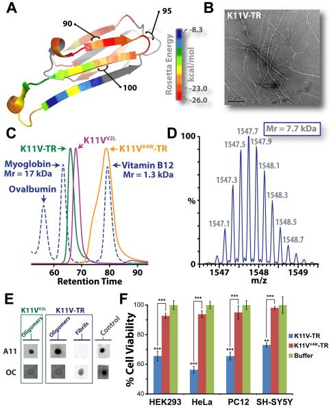
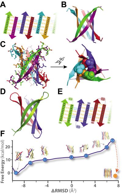
Amyloid diseases, including Alzheimer's, Parkinson's, and the prion conditions, are each associated with a particular protein in fibrillar form. These amyloid fibrils were long suspected to be the disease agents, but evidence suggests that smaller, often transient and polymorphic oligomers are the toxic entities. Here, we identify a segment of the amyloid-forming protein αB crystallin, which forms an oligomeric complex exhibiting properties of other amyloid oligomers: β-sheet-rich structure, cytotoxicity, and recognition by an oligomer-specific antibody. The x-ray-derived atomic structure of the oligomer reveals a cylindrical barrel, formed from six antiparallel protein strands, that we term a cylindrin. The cylindrin structure is compatible with a sequence segment from the β-amyloid protein of Alzheimer's disease. Cylindrins offer models for the hitherto elusive structures of amyloid oligomers.


Figures

Fig. 1
Fig. 1
The cylindrins derived from alphaB crystallin (ABC), an amyloid-forming protein, exhibit the properties of oligomeric state, immunoreactivity, and cytotoxicity commonly ascribed to small amyloid oligomers. (A) Ribbon diagram of a single subunit of ABC (16), colored by propensity to form amyloid, with red being the highest and blue the lowest propensity. The segment from residue 90 to 100, termed K11V, forms the cylindrin. (B) Representative electron micrograph of amyloid fibrils formed by the tandem repeat V2L variant of K11V, K11V-TR. (C) Overlaid size exclusion chromatograms showing protein standards (blued dashed curve) and cylindrin segments. K11VV2L (purple curve; 1.2 kDa) and K11V-TR (green curve; 2.5 kDa) cylindrin segments migrate as oligomeric complexes. A mutant form of K11V-TR that disrupts oligomer formation of the cylindrin peptide, K11VV4W-TR (orange curve; 2.7 kDa), migrates as a dimeric/monomeric species. (D) Native nanoelectrospray mass spectrum of K11V-TR peak fractions from SEC-HPLC reveals trimeric tandem repeat cylindrin oligomers, confirming that the oligomeric complexes coincide in mass with the crystallized cylindrins. Expansion of the most abundanti on series of a +5 charge state correpsonding to a molecular mass of the three K11V-TR, coinciding with the crystallographic trimeric oligomer with a mass accuracy of 3.93 ppm is shown, with m/z labels. (E) Immuno-dot blot analysis of solutions of K11VV2L and K11V-TR, and K11V-TR fibrils with prefibrillar oligomer-specific, polyclonal antibody, A11 (5), and a mixture of fibril-specific monoclonal antibodies, OC (11). Solutions of cylindrin-forming segments are recognized by A11, whereas not by the OC antibody. In contrast, K11V-TR fibrils are recognized only by the OC antibody. Positive controls are shown to the right (5). (F) Cylindrin K11V-TR is toxic to four mammalian cells lines. Cell viability levels return to nearly 100% when we tested the control variant K11VV4W-TR. All samples were at a finalconcentration of 100 μM. Results represent mean ± SEM. Student's t-test (N = 4): **, P < 0.01; and ***,P < 0.001.
Fig. 2
Fig. 2
Crystal structures of cylindrins and computed free energy change of the simulated structural transition from cylindrin to a fibril. Each colored beta-strand (arrow) is composed of eleven amino acid residues from ABC of sequence KVKVLGDVIEV (K11V). (A) Schematic of unrolled cylindrin (outside view), illustrating strand-to-strand registration. Hydrogen bonds between the main chains of neighboring strands are shown by yellow dashed lines; hydrogen bonds mediated by water bridges or side chains are shown by blue dashed lines. (B) Ribbon representation of the cylindrin crystal structure. Pairs of strands form anti-parallel dimers, which assemble around a three-fold axis down the barrel axis of the cylindrin. The height of the cylindrin is 22 Å. The inner dimension of the cylindrin, around the waist from Cα to Cα, is 12 Å, and at the splayed ends is 22 Å. (C) The cylindrin with sidechains shown as atoms, and hydrogenbonds in yellow. Twelve backbone hydrogen bonds stabilize the strong interface between tightlytwisted anti-parallel strands (e.g. between green and purple chains). The weaker interface between the pairs of tightly twisted strands is formed by four main-chain hydrogen bonds, with an additional two hydrogen bonds coming from a water bridge and two hydrogen bonds from side chain interactions (e.g. between purple and blue chains). The dry interior of the cylinder is closed by triplets of Val residues, shown as spheres, at the top and bottom. (D) Crystal structure of K11V-TR formed by three chains of 25residues each. (E) Schematic of unrolled K11V-TR cylindrin (outside view). Similar hydrogen bondingpatterns are formed as in (A). (F) The computed Gibbs free energy at 300K for a cylindrin forced to afibril. The reaction coordinate (ΔRMSD) measures the difference in root-mean-squared deviation fromthe two end points: the cylindrin and the in-register anti-parallel beta-sheet (IAB). The cylindrin set thefree energy minimum (point 1). The transition was initiated by disrupting the weak interface (points 2–3). As the cylindrin unrolls, the weak interface requires complete dissociation of backbone hydrogen bonds (points 4–5), whereas the strong interfaces maintains hydrogen bonding (point 6). The IAB has a higher free energy than the cylindrin (point 7), and when two associate and interdigitate to form a steric-zipper (point 8) the free energy drops to 5.2 kcal/mol/peptide lower than the cylindrin (Table S4).

Comment in

References

    1. Caughey B, Lansbury PT. Annu Rev Neurosci. 2003;26:267. - PubMed
    1. Xue WF, et al. J Biol Chem. 2009 Dec 4;284:34272. - PMC - PubMed
    1. Kodali R, Wetzel R. Curr Opin Struct Biol. 2007 Feb;17:48. - PubMed
    1. Kirkitadze MD, Bitan G, Teplow DB. J Neurosci Res. 2002 Sep 1;69:567. - PubMed
    1. Kayed R, et al. Science. 2003 Apr 18;300:486. - PubMed

Publication types