. 2012 May 4;287(19):15205-18.
doi: 10.1074/jbc.M111.338665. Epub 2012 Mar 7.

Role for two conserved intermembrane space proteins, Ups1p and Ups2p, [corrected] in intra-mitochondrial phospholipid trafficking

Affiliations

Role for two conserved intermembrane space proteins, Ups1p and Ups2p, [corrected] in intra-mitochondrial phospholipid trafficking

Yasushi Tamura et al. J Biol Chem. .

Erratum in

  • J Biol Chem. 2012 Aug 10;287(33):27450

Abstract

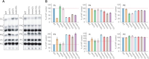
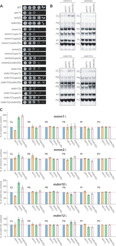
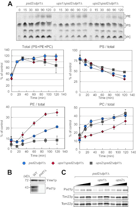
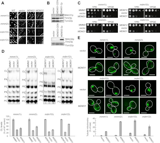
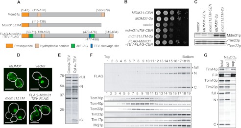
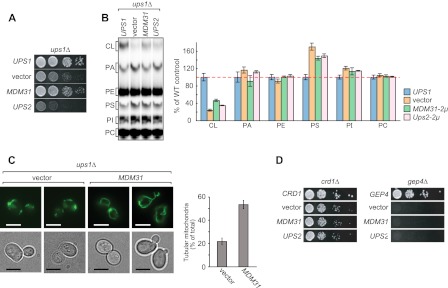
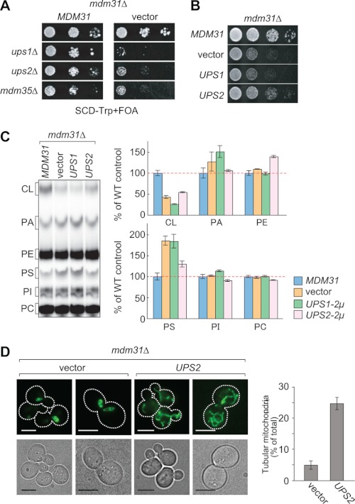
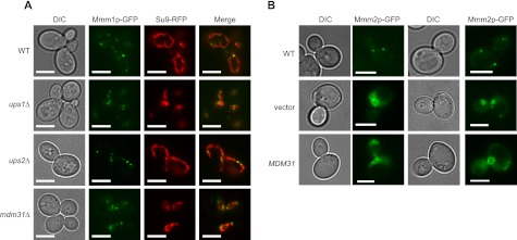
Mitochondrial membranes maintain a specific phospholipid composition. Most phospholipids are synthesized in the endoplasmic reticulum (ER) and transported to mitochondria, but cardiolipin and phosphatidylethanolamine are produced in mitochondria. In the yeast Saccharomyces cerevisiae, phospholipid exchange between the ER and mitochondria relies on the ER-mitochondria encounter structure (ERMES) complex, which physically connects the ER and mitochondrial outer membrane. However, the proteins and mechanisms involved in phospholipid transport within mitochondria remain elusive. Here, we investigated the role of the conserved intermembrane space proteins, Ups1p and Ups2p, and an inner membrane protein, Mdm31p, in phospholipid metabolism. Our data show that loss of the ERMES complex, Ups1p, and Mdm31p causes similar defects in mitochondrial phospholipid metabolism, mitochondrial morphology, and cell growth. Defects in cells lacking the ERMES complex or Ups1p are suppressed by Mdm31p overexpression as well as additional loss of Ups2p, which antagonizes Ups1p. Combined loss of the ERMES complex and Ups1p exacerbates phospholipid defects. Finally, pulse-chase experiments using [(14)C]serine revealed that Ups1p and Ups2p antagonistically regulate conversion of phosphatidylethanolamine to phosphatidylcholine. Our results suggest that Ups proteins and Mdm31p play important roles in phospholipid biosynthesis in mitochondria. Ups proteins may function in phospholipid trafficking between the outer and inner mitochondrial membranes.


Figures

FIGURE 1.
FIGURE 1.
Altered phospholipid composition in mitochondria lacking Ups proteins or ERMES subunits. A, indicated yeast cells were cultivated in YPD in the presence of 32Pi. Phospholipids were extracted from crude mitochondrial fractions and separated by TLC. PI, phosphatidylinositol. B, amounts of each lipid relative to total phospholipids were determined, and those detected in (WT) mitochondria were set to 100% (red dashed lines). Values are mean ± S.E. (n = 3).
FIGURE 2.
FIGURE 2.
Loss of Ups2p improves cell growth and CL levels in ERMES-deleted cells. A, serial dilutions of the indicated yeast cells were spotted onto YPD and cultivated at 30 °C for 2 days. B, indicated yeast cells were cultivated in YPD in the presence of 32Pi. Phospholipids were extracted from crude mitochondria fractions and analyzed by TLC. C, amounts of each lipid relative to total phospholipids were determined, and those detected in ERMES-deleted mitochondria were set to 100% (e.g. mmm1Δ, mmm2Δ, mdm10Δ, or mdm12Δ). Values are mean ± S.E. (n = 3). PI, phosphatidylinositol.
FIGURE 3.
FIGURE 3.
Loss of Ups1p and Ups2p affects conversion of PE to PC. A, cells were pulse-labeled with [14C]serine for 15 min and further cultivated for the indicated periods of time. Total phospholipids were extracted from the cells and analyzed by TLC followed by radioimaging. Total amounts of PS, PE, and PC at 15 min were set to 100% (Total). The ratio of PS, PE, or PC relative to the total amounts was determined at each time point. Values are means ± S.E. (n = 3). B, whole cell extracts were prepared from wild-type and psd1Δ cells and analyzed by immunoblotting with antibodies against Yme1p and Psd1p. C, mitochondria were isolated from the indicated cells and analyzed by immunoblotting with antibodies to Psd1p, Tim23p, and Tom22p.
FIGURE 4.
FIGURE 4.
Overexpression of Mdm31p improves cell growth, CL abundance, and mitochondrial morphology in ERMES deletion cells. A, mmm1Δ, mmm2Δ, mdm10Δ, and mdm12Δ cells with URA3-single copy plasmid harboring the corresponding gene were co-transformed with TRP1 multicopy vector harboring MDM31 or MDM32 or an empty vector. Transformants were streaked onto SCD-Trp + FOA plates and cultivated for 3 days to remove the URA3 plasmid. B, whole cell extracts were prepared from mdm31Δ (upper panel) or mdm32Δ cells (lower panel) and analyzed by immunoblotting with the indicated antibodies. Cells were transformed with single copy (CEN) and multicopy (2μ) plasmids expressing Mdm31p or Mdm32p. An empty vector was used as a negative control. C, serial dilutions of the indicated yeast cells were spotted onto YPD and YPGE, and grown for 2 and 5 days, respectively. D, ERMES-deleted cells with either the single copy plasmid harboring the corresponding gene (MMM1, MMM2, MDM10, or MDM12), an empty vector (vector), or the multicopy plasmid harboring MDM31 (MDM31) were grown in YPD in the presence of 32Pi. Phospholipids were extracted from crude mitochondrial fractions and analyzed by TLC. Amounts of CL relative to total phospholipids were determined, and those detected in mitochondria were isolated from ERMES-deleted cells with the wild-type plasmid (e.g. MMM1, MMM2, MDM10, or MDM12) were set to 100%. E, mitochondria were visualized in the indicated cells using matrix-targeted Su9-GFP. Cells were grown to log phase in YPD and examined by fluorescence microscopy and differential interference microscopy. Cells containing tubular mitochondria were scored. Values are mean ± S.E. (n = 3). At least 100 cells were examined in each experiment. Bar, 5 μm.
FIGURE 5.
FIGURE 5.
Analysis of the membrane topology of Mdm31p. A, to analyze the function of the second hydrophobic region of Mdm31p, this region was deleted (Mdm31pΔTM). To determine membrane topology of Mdm31p, 3×FLAG tags were introduced at two different locations along the protein. One tag was inserted after the presequence, and the second was placed between the first and second hydrophobic domains. In addition, a tobacco etch virus (TEV)-cleavage site was introduced before the second 3×FLAG tag (FLAG-Mdm31p-TEV-FLAG). B, serial dilution of mdm31Δ cells carrying the single (CEN) or multicopy (2μ) plasmid expressing Mdm31p, Mdm31pΔTM (mdm31ΔTM), FLAG-Mdm31p-TEV-FLAG (FLAG-MDM31-TEV-FLAG), or an empty vector (vector) were spotted onto YPD and incubated for 1 day. C, whole cells extracts prepared from the cells indicated in B were analyzed by immunoblotting with the indicated antibodies. D, mitochondria were visualized by matrix-targeted Su9-GFP in mdm31Δ cells expressing different forms of Mdm31p. Mdm31pΔTM (mdm31ΔTM) was expressed from the multicopy plasmid. E, whole cell extracts from cells expressing FLAG-Mdm31p-TEV-FLAG and either Cox4p-TEV or Cyt c1-TEV were analyzed by immunoblotting using anti-FLAG antibodies. F, mitochondria were isolated from cells expressing FLAG-Mdm31p-TEV-FLAG and Cyt c1-TEV. OM and IM vesicles were generated by osmotic shock followed by sonication. These vesicles were separated by sucrose density gradient centrifugation. Proteins from each fraction were analyzed by immunoblotting using antibodies against the indicated proteins. G, isolated mitochondria expressing FLAG-Mdm31p-TEV-FLAG and Cyt c1-TEV were treated with 0.1 m Na2CO4, and the supernatant and pellet fractions were separated by ultracentrifugation. Proteins were analyzed by immunoblotting using antibodies against the indicated proteins. C and N, C- and N-terminal halves of Mdm231p.
FIGURE 6.
FIGURE 6.
Mdm31p overexpression ameliorates cell growth, CL amounts, and mitochondrial morphology in ups1Δ cells. A, serial dilutions of ups1Δ cells with single copy plasmids expressing UPS1 (UPS1), empty vector (vector), multicopy plasmids expressing Mdm31p (MDM31), or Ups2p (UPS2) were spotted onto YPD and cultivated at 30 °C for 2 days. B, yeast cells used in A were grown in YPD in the presence of 32Pi. Phospholipids were extracted from crude mitochondrial fractions and separated by TLC. Amounts of each lipid relative to total phospholipids were determined, and those detected in mitochondria from ups1Δ cells containing the Ups1p plasmid were set to 100% (e.g. UPS1). Values are mean ± S.E. (n = 3). PI, phosphatidylinositol. C, ups1Δ cells containing the multicopy plasmid harboring MDM31 (MDM31) or an empty vector (vector) were grown in YPD to log phase. Mitochondria were visualized using matrix-targeted Su9-GFP. Cells containing tubular mitochondria were scored. Values are mean ± S.E. (n = 3). At least 100 cells were examined in each experiment. Bar, 5 μm. D, serial dilutions of crd1Δ and gep4Δ cells carrying the multicopy plasmid harboring MDM31 (MDM31) or UPS2 (UPS2), or an empty vector (vector), were spotted onto YPD and grown for 2 days.
FIGURE 7.
FIGURE 7.
Ups2p overexpression partially rescues defects in cell growth, CL levels, and mitochondrial morphology in mdm31Δ cells. A, UPS1, UPS2, or MDM35 gene was deleted in mdm31Δ cells carrying the URA3 single copy plasmid harboring the MDM31 gene. The resulting yeast cells were transformed with the TRP1 single copy plasmid harboring MDM31 or a TRP1 empty vector. These cells were spotted onto the 5′-FOA-containing SCD-Trp medium and incubated for 2 days. B, serial dilutions of mdm31Δ cells carrying the single copy plasmid expressing Mdm31p (MDM31), the multicopy plasmids expressing Ups1p (UPS1) or Ups2p (UPS2), or an empty vector (vector) were spotted onto YPD and grown for 2 days. C, yeast cells used in B were grown in YPD in the presence of 32Pi. Phospholipid was extracted from crude mitochondrial fractions and analyzed by TLC. Amounts of each lipid were quantified and normalized to those in mdm31Δ mitochondria expressing Mdm31p. Values are mean ± S.E. (n = 3). PI, phosphatidylinositol. D, mitochondrial morphology in mdm31Δ cells carrying the multicopy plasmid harboring UPS2 (UPS2) or an empty vector (vector) were visualized using Su9-GFP. Cells were cultivated in YPD to log prior to observation. Cells containing tubular mitochondria were scored. Values are means ± S.E. (n = 3). At least 100 cells were examined in each experiment. Bar, 5 μm.
FIGURE 8.
FIGURE 8.
Ups1p, Ups2p, and Mdm31p are not required for formation of ER-mitochondria contact sites. A, GFP-tagged Mmm1p (Mmm1p-GFP) and mitochondria-targeted RFP (Su9-RFP) were expressed in wild-type, ups1Δ, ups2Δ, and mdm31Δ cells. B, GFP-tagged Mmm2p (Mmm2p-GFP) was expressed from plasmid, pMY3 (33), in mdm12Δ cells with the multicopy plasmid expressing Mdm31p or an empty vector. Bar, 5 μm; DIC, differential interference contrast.

Similar articles

Cited by

References

    1. Claypool S. M. (2009) Cardiolipin, a critical determinant of mitochondrial carrier protein assembly and function. Biochim. Biophys. Acta 1788, 2059–2068 - PMC - PubMed
    1. Joshi A. S., Zhou J., Gohil V. M., Chen S., Greenberg M. L. (2009) Cellular functions of cardiolipin in yeast. Biochim. Biophys. Acta 1793, 212–218 - PMC - PubMed
    1. Jiang F., Ryan M. T., Schlame M., Zhao M., Gu Z., Klingenberg M., Pfanner N., Greenberg M. L. (2000) Absence of cardiolipin in the crd1 null mutant results in decreased mitochondrial membrane potential and reduced mitochondrial function. J. Biol. Chem. 275, 22387–22394 - PubMed
    1. van der Laan M., Meinecke M., Dudek J., Hutu D. P., Lind M., Perschil I., Guiard B., Wagner R., Pfanner N., Rehling P. (2007) Motor-free mitochondrial presequence translocase drives membrane integration of preproteins. Nat. Cell Biol. 9, 1152–1159 - PubMed
    1. Kutik S., Rissler M., Guan X. L., Guiard B., Shui G., Gebert N., Heacock P. N., Rehling P., Dowhan W., Wenk M. R., Pfanner N., Wiedemann N. (2008) The translocator maintenance protein Tam41 is required for mitochondrial cardiolipin biosynthesis. J. Cell Biol. 183, 1213–1221 - PMC - PubMed

Publication types

MeSH terms