. 2012;7(3):e31719.
doi: 10.1371/journal.pone.0031719. Epub 2012 Mar 5.

Detecting remote sequence homology in disordered proteins: discovery of conserved motifs in the N-termini of Mononegavirales phosphoproteins

Affiliations

Detecting remote sequence homology in disordered proteins: discovery of conserved motifs in the N-termini of Mononegavirales phosphoproteins

David Karlin et al. PLoS One. 2012.

Abstract

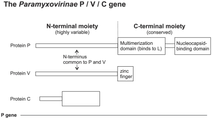
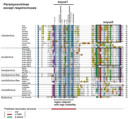
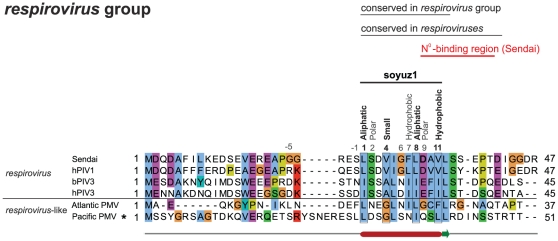
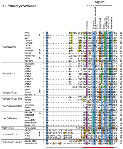
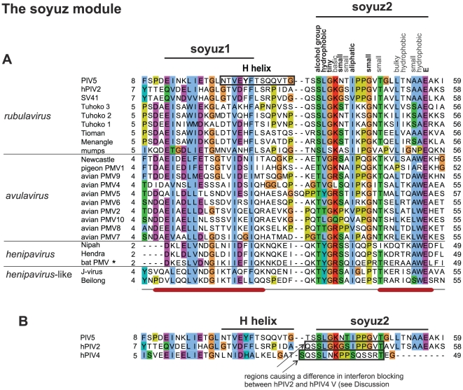
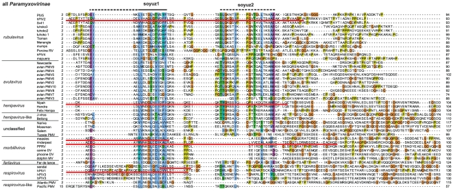
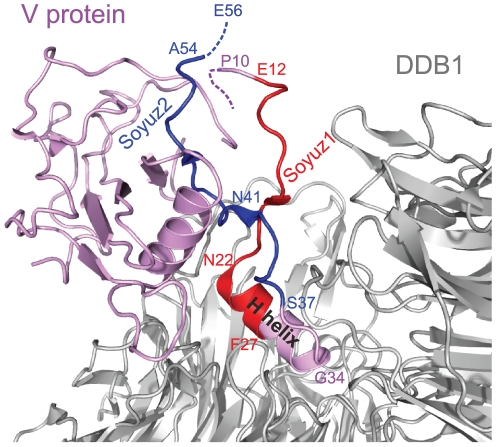
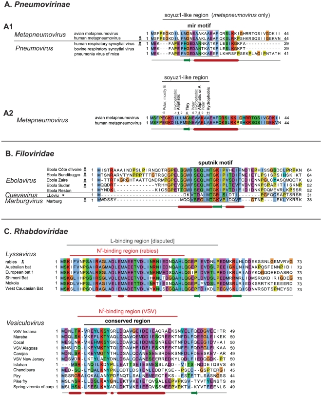
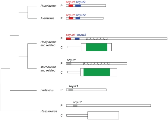
Paramyxovirinae are a large group of viruses that includes measles virus and parainfluenza viruses. The viral Phosphoprotein (P) plays a central role in viral replication. It is composed of a highly variable, disordered N-terminus and a conserved C-terminus. A second viral protein alternatively expressed, the V protein, also contains the N-terminus of P, fused to a zinc finger. We suspected that, despite their high variability, the N-termini of P/V might all be homologous; however, using standard approaches, we could previously identify sequence conservation only in some Paramyxovirinae. We now compared the N-termini using sensitive sequence similarity search programs, able to detect residual similarities unnoticeable by conventional approaches. We discovered that all Paramyxovirinae share a short sequence motif in their first 40 amino acids, which we called soyuz1. Despite its short length (11-16aa), several arguments allow us to conclude that soyuz1 probably evolved by homologous descent, unlike linear motifs. Conservation across such evolutionary distances suggests that soyuz1 plays a crucial role and experimental data suggest that it binds the viral nucleoprotein to prevent its illegitimate self-assembly. In some Paramyxovirinae, the N-terminus of P/V contains a second motif, soyuz2, which might play a role in blocking interferon signaling. Finally, we discovered that the P of related Mononegavirales contain similarly overlooked motifs in their N-termini, and that their C-termini share a previously unnoticed structural similarity suggesting a common origin. Our results suggest several testable hypotheses regarding the replication of Mononegavirales and suggest that disordered regions with little overall sequence similarity, common in viral and eukaryotic proteins, might contain currently overlooked motifs (intermediate in length between linear motifs and disordered domains) that could be detected simply by comparing orthologous proteins.


Conflict of interest statement

Competing Interests: The authors have declared that no competing interests exist.

Figures

Figure 1
Figure 1. Organization of the Paramyxovirinae P gene.
The P, V and C proteins are encoded from alternative reading frames. V is produced in all Paramyxovirinae genera whereas C is only produced in henipaviruses, morbilliviruses, and respiroviruses.
Figure 2
Figure 2. Alignment of the N-termini of P from all Paramyxovirinae except respiroviruses (see Figure 3 ), realized with MAFFT and coloured according to the ClustalX scheme .
Abbreviations and accession numbers are in Table 1. Positions with conserved physico-chemical character are indicated above the alignment, in bold if the character is strictly conserved (100%) and in normal font if it is generally conserved (>80%). Numbering of the soyuz1 motif (above the alignment) starts at the first strictly conserved position. Unpublished sequences are shown by an asterisk.
Figure 3
Figure 3. Alignment of the N-termini of P from respiroviruses.
Positions matching the soyuz1 of the other Paramyxovirinae are indicated above the alignment (see Figure 2). An experimentally characterized substitution in Sendai virus is in bold.
Figure 4
Figure 4. Alignment of the N-termini of P from all Paramyxovirinae.
Conventions as in Figure 2. The part of soyuz1 not conserved in respiroviruses is indicated by a dashed line above the alignment. Species pathogenic for humans are marked by a skull and crossbones. Experimentally characterized substitutions in measles virus and Sendai virus are in bold.
Figure 5
Figure 5. N-termini of P from the rubulaviruses, avulaviruses, and henipaviruses that have the soyuz2 motif.
Conventions as in Figure 2. (A) Experimentally characterized substitutions in soyuz2 and in the H helix are in bold. (B) Comparison of the N-termini of the V protein of PIV5 and hPIV2 (which both have a soyuz2 motif) with that of hPIV4 (which lacks the soyuz2 motif).
Figure 6
Figure 6. Alignment of the first 100aa of all Paramyxovirinae P. Conventions as in Figure 2 .
The boundaries of N°-binding regions (underlined in red) have generally been determined indirectly (Table 3), and thus should be taken as approximate. Regions downstream of soyuz1 and soyuz2 (90–330aa in length, of which only ∼50aa are visible on the figure) are unalignable between different genera of Paramyxovirinae.
Figure 7
Figure 7. Structure of the V protein from parainfluenza virus 5 bound to DDB1.
The PDB accession number of the structure is 2HYE. Aa 1–9 and aa 55–80 of V, encompassing the last 2aa of soyuz2, are not visible in the crystal structure, presumably because they are disordered (see text). Soyuz1 is coloured red and soyuz2 blue. The H helix of V, bound to DDB1, is indicated; it partially overlaps with soyuz1.
Figure 8
Figure 8. Alignments of the N-termini of P from Pneumovirinae, Filoviridae and Rhabdoviridae.
Conventions as in Figure 2. Abbreviations and accession numbers are in Table 2. (A) Mir motif of Pneumovirinae. A1 – Alignment of the N-terminus of P of both metapneumoviruses and pneumoviruses. A2 – Same alignment as in A1 but restricted to metapneumoviruses. Positions corresponding to soyuz1 are indicated above the alignment. The coloring of sequence conservation is different from A1 since conservation is now based only on the two metapneumovirus sequences. (B) Sputnik motif of Filoviridae. The asterisk indicates the newly published sequence of LLoviu virus. (C) N-termini of the P of two Rhabdoviridae genera: lyssavirus and vesiculovirus. A disputed L-binding site in lyssavirus P is indicated . The boundaries of the N°-binding region of VSV P were obtained from the crystal structure of N°-P .
Figure 9
Figure 9. Structural superposition of the C-termini of two Paramyxovirinae and Filoviridae P.
FATCAT superposition between the measles virus X domain (PDB accession number 1T60, chain A), in red, and the Zaire ebolavirus IID domain (3FKE, chain A), in green. N and C refer to N- and C-termini.
Figure 10
Figure 10. Regions with sequence similarity in Paramyxovirinae P and C.
The N-termini of Paramyxovirinae P and the C proteins that overlap them are represented to scale (the N-terminus of henipavirus P is about 380aa long). The phylogenetic relationships between different genera are shown on the left as a cladogram based on . Regions with statistically significant similarity (and thus homologous) are shown in the same colours, whereas regions that have subsignificant similarity are shown in grey. The crisscrossed regions of henipavirus and morbillivirus P are homologous, even though they have no detectable similarity, since they overlap homologous regions of C, in green (see Discussion).

References

    1. Whelan SP, Barr JN, Wertz GW. Transcription and replication of nonsegmented negative-strand RNA viruses. Current Topics in Microbiology and Immunology. 2004;283:61–119. - PubMed
    1. Fontana JM, Bankamp B, Rota PA. Inhibition of interferon induction and signaling by paramyxoviruses. Immunological Reviews. 2008;225:46–67. - PubMed
    1. Habchi J, Longhi S. Structural disorder within paramyxovirus nucleoproteins and phosphoproteins. Molecular Biosystems. 2011;8:69–81. - PubMed
    1. Leyrat C, Gerard FCA, Ribeiro ED, Ivanov I, Ruigrok RWH, et al. Structural disorder in proteins of the rhabdoviridae replication complex. Protein and Peptide Letters. 2010;17:979–987. - PubMed
    1. Karlin D, Ferron F, Canard B, Longhi S. Structural disorder and modular organization in paramyxovirinae N and P. Journal of General Virology. 2003;84:3239–3252. - PubMed

Publication types