. 2012;7(3):e32049.
doi: 10.1371/journal.pone.0032049. Epub 2012 Mar 5.

Aloe vera induced biomimetic assemblage of nucleobase into nanosized particles

Affiliations

Aloe vera induced biomimetic assemblage of nucleobase into nanosized particles

Arun Chauhan et al. PLoS One. 2012.

Abstract

Aim: Biomimetic nano-assembly formation offers a convenient and bio friendly approach to fabricate complex structures from simple components with sub-nanometer precision. Recently, biomimetic (employing microorganism/plants) synthesis of metal and inorganic materials nano-particles has emerged as a simple and viable strategy. In the present study, we have extended biological synthesis of nano-particles to organic molecules, namely the anticancer agent 5-fluorouracil (5-FU), using Aloe vera leaf extract.

Methodology: The 5-FU nano- particles synthesized by using Aloe vera leaf extract were characterized by UV, FT-IR and fluorescence spectroscopic techniques. The size and shape of the synthesized nanoparticles were determined by TEM, while crystalline nature of 5-FU particles was established by X-ray diffraction study. The cytotoxic effects of 5-FU nanoparticles were assessed against HT-29 and Caco-2 (human adenocarcinoma colorectal) cell lines.

Results: Transmission electron microscopy and atomic force microscopic techniques confirmed nano-size of the synthesized particles. Importantly, the nano-assembled 5-FU retained its anticancer action against various cancerous cell lines.

Conclusion: In the present study, we have explored the potential of biomimetic synthesis of nanoparticles employing organic molecules with the hope that such developments will be helpful to introduce novel nano-particle formulations that will not only be more effective but would also be devoid of nano-particle associated putative toxicity constraints.


Conflict of interest statement

Competing Interests: The authors have declared that no competing interests exist.

Figures

Figure 1
Figure 1. Spectrophotometric analysis of 5-FU nano-particles.
(A) Time kinetic effect of Aloe vera leaf extract mediated synthesis of 5-FU nano-particles employing UV Spectrophotometric analysis. The 5-FU solution was incubated with Aloe vera extract (5 ml of 10−3 M 5-FU and 5 ml Aloe vera extract; final volume of reaction mixture 10 ml) for specified time period (0–24 h). The incubation mixture was scanned in UV range to analyze characteristic peaks. The spectrum shown at zero time point actually corresponds to 5-FU nanoparticles obtained instantly just after initial mixing of free 5-FU with Aloe vera leaf extract. (B) Concentration dependent kinetics of Aloe vera leaf extract mediated synthesis of 5-FU nano-particles as revealed by UV Spectrophotometric analysis. UV spectra of 5-FU nano-particles synthesized using increasing amounts of Aloe vera extract. (C) Emission spectra of 5-FU nano-particles synthesized using Aloe vera extract. The 5-FU solution (10−3 M) was incubated with increasing volume of Aloe vera extract and scanned employing spectrofluorimeter. The excitation wave length was 260 nm.
Figure 2
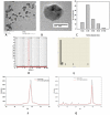
Figure 2. Transmission electron microscopic images of 5-FU nanoparticles.
(A) Representative TEM image of 5-FU nanoparicles synthesized by incubating 5 ml of 10−3 M 5-FU solution with 5 ml of Aloe vera extract for 48 h. (B) TEM image of the plate like hexagonal 5-FU nano-particles prepared from 5-FU (10−3 M) with Aloe vera leaf extract (5 ml). HRTEM image shows different section of hexagonal nano-particles of 5-FU. (C) Variation in particle size diameter upon incubation of 10−3 M 5-FU solution (5 ml) with 5 ml Aloe vera extract for 48 h. (D) Particle size analysis of 5-FU nano-particles as determined by Nanofox particle analyzer. (E) Graph showing size of the nano-particle as obtained by Dynamic Light Scattering studies. (F–G) The X-ray diffraction analysis of 5-FU nano-particles. The X-ray diffraction pattern shows characteristic intense peak at diffraction angle (2θ) of 16°, 19°, 22° and 28°. X-ray diffraction of 5-FU nano-particle was analyzed at diffraction angle (2θ) of (F) 20–30 and (G) 25–30 respectively.
Figure 3
Figure 3. Atomic Force Microscopic study of 5-FU nano-particles.
Representative AFM image of 5-FU nano particles synthesized by incubation of 5 ml of 10−3 M 5-FU with 5 ml Aloe vera extract for 48 hr. (A) 2D, (B) 3D images of 5-FU nano-particles (C) Histogram representation of AFM analysis of 5-FU nano-particles.
Figure 4
Figure 4. FTIR spectra of 5-FU nano-particles.
spectrum 1; Aloe vera extract, spectrum II; 5-FU only, spectrum III; 5-FU nano-particles synthesized with 3 ml of Aloe vera extract, spectrum IV; 5-FU nano-particles synthesized with 5 ml of Aloe vera extract. All the FTIR spectra were recorded after 48 hr of incubation of 5-FU with Aloe vera leaf extract.
Figure 5
Figure 5. Release profile of 5-FU nano-particles under various conditions.
To examine the release kinetics of 5-FU nano-particles, multiple samples of the formulation were dispensed into various micro vials. To each vial, 1.0 ml of release medium (20 mM sterile PBS, serum or histidine buffer) was dispensed. After stipulated time period, the suspension was centrifuged and an aliquot was analyzed for 5-FU content spectrophotometrically as described in Methodology section.
Figure 6
Figure 6. Cytotoxic effect of 5-FU nano-particles.
Against (A) HT-29 and (B) Caco-2 cell lines. MTT assay was used to determine the differential cytotoxicity of 5-FU nano-particles against both cell lines. Cells were dispensed at the density of 5×10⁴ cells per well in U-bottom 96 well plates in triplicate, and treated with increasing concentration of various 5-FU nano formulations. After 48 h of incubation, 20 µl of MTT reagent was added and plate was further incubated for 48 h. It was followed by addition of 100 µl of lysis buffer. The plates were further incubated for 2 hr and absorbance was read at 570 nm.
Figure 7
Figure 7. Effect of 5-FU nano-particles on expression of pro/anti apoptotic factors in Caco-2 cell line.
Cell lysate was prepared as described in Methodology and analyzed for protein expression using specific antibodies. To quantify equal loading, the membranes were also probed with β-actin antibody. The intensity of bands was quantified using image analysis software on an image gel documentation system. Lane 1, RPMI only; Lane 2, Aloe vera leaf extract only; Lane 3, 5-FU only; Lane 4, 5-FU nano-particles prepared by mixing of 5-FU (10−3 M) solution with 3 ml of Aloe vera extract and Lane 5, 5-FU nano-particles prepared by mixing of 5-FU (10−3 M) solution with 5 ml of Aloe vera extract.
Figure 8
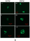
Figure 8. Involvement of caspase-9 in 5-FU mediated apoptosis of Caco-2 cells as revealed by confocal microscopy.
Caco-2 cell, were treated with (a) RPMI only, (b) free 5-FU drug, and (c) 5-FU nano-particles for 24 h. After incubation for stipulated time period with various 5-FU preparations, cells were washed and fixed with 4% paraformaldehyde and 0.19% picric acid in PBS (pH 7.4) for 1 h at room temperature (RT). Fixed cells were permeabilized with 0.1% SDS in PBS at RT for 10 min, blocked with 2% FCS, stained with a polyclonal antibody which detects only cleaved 35 kDa caspase-9 (BD, India). The interaction was revealed employing a conjugated goat anti-rabbit IgG-FITC probe (Sigma, India). The cells were then visualized under a fluorescence microscope (excitation at 488 nm emission at 505–530 nm.) scale bar, 10 µm.
Figure 9
Figure 9. Ball and socket model of 5-FU nano-particles.
(A) Proposed structure of 5-FU nano-particles, (B) Simulated molecular model of 5-FU nanoparticle assembly using ChemDraw software.

Similar articles

Cited by

References

    1. Roco MC. Nanotechnology: convergence with modern biology and medicine. Curr Opin Biotechnol. 2003;14:337–343. - PubMed
    1. Gupta RB, Kompella Nanoparticle Technology for Drug Delivery. 2006. pp. 1–379. Taylor & Francis Group, New York, U.B. Eds.
    1. Wilkinson JM. Nanotechnology applications in medicine. Med Device Technol. 2003;14:29–31. - PubMed
    1. Schimidt J, Montemagno C. Using machines in cells Drug Discov. Today. 2002;7:500–503. - PubMed
    1. Lobenberg R, Kreuter J. Macrophage targeting of azidothymidine: a promising strategy for AIDS therapy. AIDS Res Hum Retroviruses. 1996;12:1709–1715. - PubMed

Publication types