. 2012;7(3):e32500.
doi: 10.1371/journal.pone.0032500. Epub 2012 Mar 5.

eIF2α kinases regulate development through the BzpR transcription factor in Dictyostelium discoideum

Affiliations

eIF2α kinases regulate development through the BzpR transcription factor in Dictyostelium discoideum

Charles K Singleton et al. PLoS One. 2012.

Abstract

Background: A major mechanism of translational regulation in response to a variety of stresses is mediated by phosphorylation of eIF2α to reduce delivery of initiator tRNAs to scanning ribosomes. For some mRNAs, often encoding a bZIP transcription factor, eIF2α phosphorylation leads to enhanced translation due to delayed reinitiation at upstream open reading frames. Dictyostelium cells possess at least three eIF2α kinases that regulate various portions of the starvation-induced developmental program. Cells possessing an eIF2α that cannot be phosphorylated (BS167) show abnormalities in growth and development. We sought to identify a bZIP protein in Dictyostelium whose production is controlled by the eIF2α regulatory system.

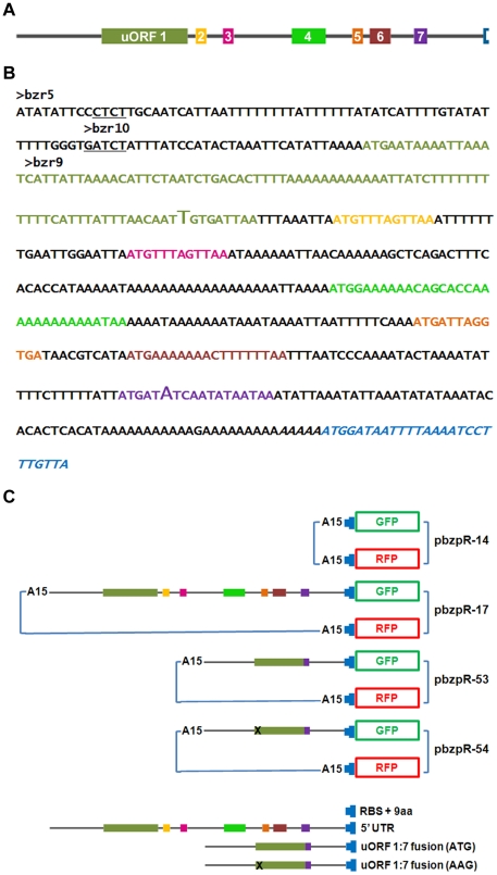
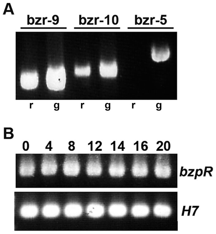
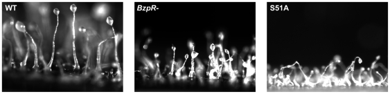
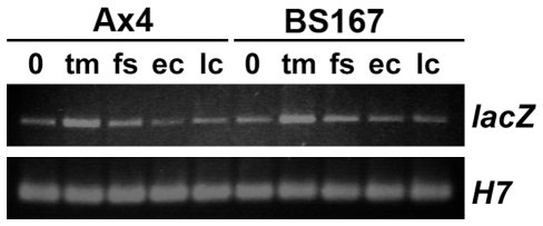
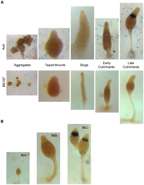
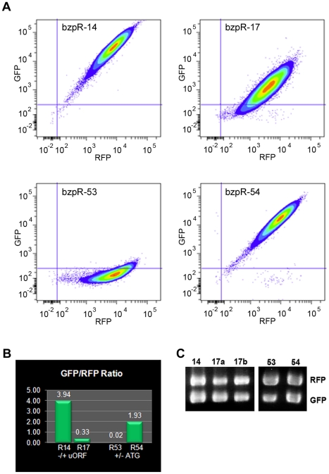
Principal findings: Cells disrupted in the bzpR gene had similar developmental defects as BS167 cells, including small entities, stalk defects, and reduced spore viability. β-galactosidase production was used to examine translation from mRNA containing the bzpR 5' UTR. While protein production was readily apparent and regulated temporally and spatially in wild type cells, essentially no β-galactosidase was produced in developing BS167 cells even though the lacZ mRNA levels were the same as those in wild type cells. Also, no protein production was observed in strains lacking IfkA or IfkB eIF2α kinases. GFP fusions, with appropriate internal controls, were used to directly demonstrate that the bzpR 5' UTR, possessing 7 uORFs, suppressed translation by 12 fold. Suppression occurred even when all but one uORF was deleted, and translational suppression was removed when the ATG of the single uORF was mutated.

Conclusions: The findings indicate that BzpR regulates aspects of the development program in Dictyostelium, serving as a downstream effector of eIF2α phosphorylation. Its production is temporally and spatially regulated by eIF2α phosphorylation by IfkA and IfkB and through the use of uORFs within the bzpR 5' UTR.


Conflict of interest statement

Competing Interests: The authors have declared that no competing interests exist.

Figures

Figure 1
Figure 1. The bzpR 5′ UTR sequence and plasmids used to test translational suppression.
A. A ‘to scale’ schematic of the seven uORFs within the bzpR 5′ UTR, color coded to correspond with panels B and C. B. The bzpR 5′ UTR sequence and the first nine codons (blue) of the coding region. The 5′ ends of the three primers, bzr-9, bzr-10, and bzr-5, used in RT-PCR to localize the transcriptional start site, are indicated. Transcription likely starts between the two underlined regions. The sequence shown was fused to the GFP gene in pbzpR-17. The italicized region, consisting only of the ribosome binding site and the first 9 codons, is the no-UTR control fused to the GFP and RFP genes in pbzpR-14 and to the RFP gene in pbzpR-17, -53, and -54. The larger font T and A in the first and last uORFs represent the fusion junction in the internal deletion constructs (pbzpR-53 and -54). C. Schematic diagram of the plasmids used to demonstrate suppression of translation by the bzpR 5′ UTR. A15 represents the actin 15 promoter driving transcription of each fusion. Thin blue lines represent the remaining portions of the plasmids, including transcriptional terminators and selectable drug resistance genes.
Figure 2
Figure 2. RT-PCR localization of transcriptional start site of bzpR gene and determination of relative mRNA levels.
A. RNA and genomic DNA were isolated from growing Ax4 cells and used as templates in RT-PCR (r) or PCR (g) reactions. A 3′ primer (bzr-3) corresponding to sequences at the beginning of the coding region was used with three different 5′ primers corresponding to sequences progressively further upstream of the coding region (see Fig. 1A). Bzr-9 lies within the first uORF, bzr-10 lies just upstream of uORF 1, and bzr-5 is 41 residues further upstream of -10. B. RNA was isolated from Ax4 growing cells (0) and cells plated for development for the indicated times (in hours) and used in RT-PCR reactions with bzr-3 and -10. H7 specific primers were used as an internal control as H7 is expressed constitutively during growth and development. Conditions were optimized to reveal differences in RNA levels of two to ten-fold.
Figure 3
Figure 3. Fruiting bodies of Ax4 (wild type), BS168 (bzpR null), and BS167 (eIF2α S51A knockin).
Cells were grown in the presence of bacteria, harvested, and plated for development. Photographs were taken at 48× after the filter was adhered to a gel slice in a vertical position.
Figure 4
Figure 4. RT-PCR determination of relative levels of β-galactosidase mRNA in Ax4 and BS167 transformed with pbzpR-9.
Wild type (Ax4) and eIF2α S51A knockin (BS167) cells were grown in the presence of bacteria, harvested, and plated for development. RNA was isolated from growing cells (0) and at the tipped mound (tm), finger/slug (fs), early culminant (ec), and late culminant (lc) stages. Primers used were bzr-10 and lacZ-4. H7 primers were used in parallel reactions to confirm RNA concentrations. Conditions were optimized to reveal differences in RNA levels of two to ten-fold.
Figure 5
Figure 5. Spatial and temporal expression of β-galactosidase protein driven by the bzpR promoter and 5′ UTR.
Strains transformed with pbzpR-9 were grown in the presence of bacteria, harvested, and plated for development. At appropriate times, filters of the developing cells were fixed and stained for β-galactosidase activity. All samples were stained for 5 hours at room temperature, washed, and photographed in glycerol at 48× with the exception of tipped mounds, which were photographed at 75× to capture detail. A. Various stages are shown for Ax4 (wild type) and BS167 (eIF2α S51A knockin). B. Late culminants are shown for BS153 (ifkA null), BS160 (ifkB null), and BS166 (ifkC null).
Figure 6
Figure 6. Translational repression by the bzpR 5′ UTR.
Ax4 cells were transformed with the various bzpR 5′ UTR constructs: pbzpR-14 (no-UTR GFP, no-UTR RFP), pbzpR-17 (5′ UTR GFP, no-UTR RFP), pbzpR-53 (GFP: modified 5′ UTR with a single uORF, that being the fusion of uORF 1 and 7; no-UTR RFP), or pbzpR-54 (GFP: modified 5′ UTR mutant with the single fused uORF possessing an AAG start codon mutation; no-UTR RFP). Flow cytometry was used to compare levels of GFP and RFP in cells of each strain. A. Colored contour plots of GFP versus RFP fluorescence intensities for cells transformed with the indicated constructs. B. The GFP and RFP mean fluorescence values from the contour plots were used to determine the GFP to RFP ratios for each strain. Mean values were: pbzpR-14, 34,372 (GFP), 8735 (RFP); pbzpR-17, 1863 (GFP), 5675 (RFP); pbzR-53, 172 (GFP), 9570 (RFP); pbzpR-54, 19,193 (GFP), 9966 (RFP). C. RNA was isolated from cells transformed with each construct, and RT-PCR was carried out with RFP and GFP specific primers to compare relative levels of RFP and GFP mRNA. Two independently transformed populations are shown for pbzpR-17 (a and b). Several independently transformed populations with pbzpR-53 or pbzpR-54 gave identical results to those shown, i.e., a slight reduction in relative levels of GFP mRNA in -53.

References

    1. Dever TE. Gene-specific regulation by general translation factors. Cell. 2002;108:545–556. - PubMed
    1. Brewer JW, Diehl JA. PERK mediates cell-cycle exit during the mammalian unfolded protein response. Proc Natl Acad Sci USA. 2000;97:12625–12630. - PMC - PubMed
    1. Dever TE, Feng L, Wek RC, Cigan AM, Donahue TF, et al. Phosphorylation of initiation factor 2 alpha by protein kinase GCN2 mediates gene-specific translational control of GCN4 in yeast. Cell. 1992;68:585–596. - PubMed
    1. Fernandez J, Yaman I, Sarnow P, Snider MD, Hatzoglou M. Regulation of internal ribosomal entry site mediated translation by phosphorylation of the translation initiation factor eIF2alpha. J Biol Chem. 2002;277:19198–19205. - PubMed
    1. Harding HP, Novoa I, Zhang YH, Zeng HQ, Wek RC, et al. Regulated translation initiation controls stress-induced gene expression in mammalian cells. Mol Cell. 2000;6:1099–1108. - PubMed

Publication types

MeSH terms

LinkOut - more resources