

Differential analysis of ovarian and endometrial cancers identifies a methylator phenotype
- PMID: 22403726
- PMCID: PMC3293923
- DOI: 10.1371/journal.pone.0032941
Differential analysis of ovarian and endometrial cancers identifies a methylator phenotype
Abstract
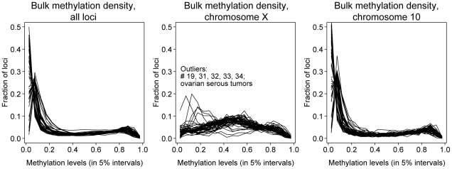
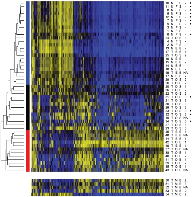
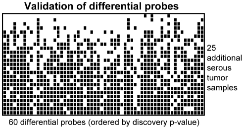
Despite improved outcomes in the past 30 years, less than half of all women diagnosed with epithelial ovarian cancer live five years beyond their diagnosis. Although typically treated as a single disease, epithelial ovarian cancer includes several distinct histological subtypes, such as papillary serous and endometrioid carcinomas. To address whether the morphological differences seen in these carcinomas represent distinct characteristics at the molecular level we analyzed DNA methylation patterns in 11 papillary serous tumors, 9 endometrioid ovarian tumors, 4 normal fallopian tube samples and 6 normal endometrial tissues, plus 8 normal fallopian tube and 4 serous samples from TCGA. For comparison within the endometrioid subtype we added 6 primary uterine endometrioid tumors and 5 endometrioid metastases from uterus to ovary. Data was obtained from 27,578 CpG dinucleotides occurring in or near promoter regions of 14,495 genes. We identified 36 locations with significant increases or decreases in methylation in comparisons of serous tumors and normal fallopian tube samples. Moreover, unsupervised clustering techniques applied to all samples showed three major profiles comprising mostly normal samples, serous tumors, and endometrioid tumors including ovarian, uterine and metastatic origins. The clustering analysis identified 60 differentially methylated sites between the serous group and the normal group. An unrelated set of 25 serous tumors validated the reproducibility of the methylation patterns. In contrast, >1,000 genes were differentially methylated between endometrioid tumors and normal samples. This finding is consistent with a generalized regulatory disruption caused by a methylator phenotype. Through DNA methylation analyses we have identified genes with known roles in ovarian carcinoma etiology, whereas pathway analyses provided biological insight to the role of novel genes. Our finding of differences between serous and endometrioid ovarian tumors indicates that intervention strategies could be developed to specifically address subtypes of epithelial ovarian cancer.
Conflict of interest statement
Figures
References
-
- Wu R, Hendrix-Lucas N, Kuick R, Zhai Y, Schwartz DR, et al. Mouse model of human ovarian endometrioid adenocarcinoma based on somatic defects in the Wnt/beta-catenin and PI3K/Pten signaling pathways. Cancer Cell. 2007;11:321–333. - PubMed
-
- Marquez RT, Baggerly KA, Patterson AP, Liu J, Broaddus R, et al. Patterns of gene expression in different histotypes of epithelial ovarian cancer correlate with those in normal fallopian tube, endometrium, and colon. Clin Cancer Res. 2005;11:6116–6126. - PubMed
-
- Folkins AK, Jarboe EA, Roh MH, Crum CP. Precursors to pelvic serous carcinoma and their clinical implications. Gynecol Oncol. 2009;113:391–396. - PubMed
Publication types
MeSH terms
Grants and funding
LinkOut - more resources
Full Text Sources
Other Literature Sources
Medical
Miscellaneous
