Review
. 2012:41:157-77.
doi: 10.1146/annurev-biophys-101211-113227. Epub 2012 Feb 23.

Bacterial mechanosensitive channels--MscS: evolution's solution to creating sensitivity in function

Affiliations
Review

Bacterial mechanosensitive channels--MscS: evolution's solution to creating sensitivity in function

James H Naismith et al. Annu Rev Biophys. 2012.

Abstract

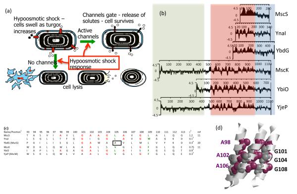
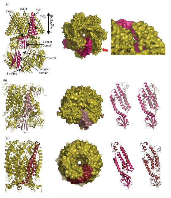
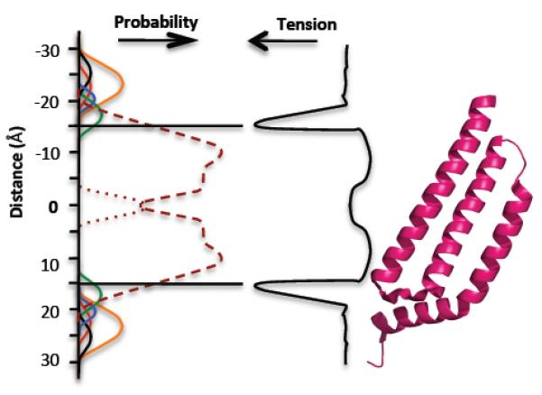
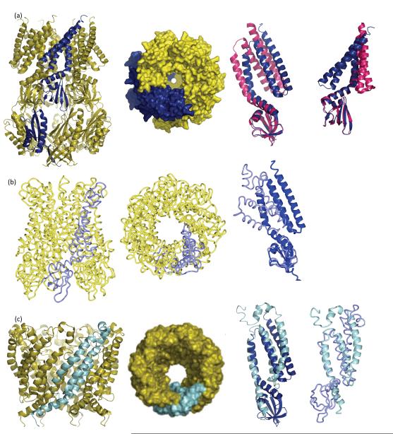
The discovery of mechanosensing channels has changed our understanding of bacterial physiology. The mechanosensitive channel of small conductance (MscS) is perhaps the most intensively studied of these channels. MscS has at least two states: closed, which does not allow solutes to exit the cytoplasm, and open, which allows rapid efflux of solvent and solutes. The ability to appropriately open or close the channel (gating) is critical to bacterial survival. We briefly review the science that led to the isolation and identification of MscS. We concentrate on the structure-function relationship of the channel, in particular the structural and biochemical approaches to understanding channel gating. We highlight the troubling discrepancies between the various models developed to understand MscS gating.


Figures

Fig 1
Fig 1
Variation and function in mechanosensitive channel of small conductance (MscS) channel homologs. (a) Mechanosensitive (MS) channels protect against cell lysis during hypoosmotic shock (downshock). During growth at high osmolarity, bacterial cells accumulate high levels of solute. Transfer to a dilute medium results in rapid inrush of water that requires the activation of the channels to elicit solute egress from the cell. Failure of channels to be activated leads to cell lysis, resulting in a number of distinct forms (left to right): fragments, viable cells, cell-like ghosts, and empty sacs (figure adapted from M. Reuter, N. Hayward, S. Miller, D. Dryden & I.R. Booth, unpublished data). (b) Homologs of MscS exhibit varying sizes, owing principally to extensions at the N terminus. The figure shows the hydrophobicity plot (Kyte-Doolittle, w = 9) (30) for the six MscS homologs in Escherichia coli. Blocks of sequence are highlighted as follows: periplasmic regions, green; membrane regions, gray; cytoplasmic domains, orange. Note that YbdG has a larger cytoplasmic domain than the other homologs due to an ~50-amino-acid insertion. (c) Comparison of the pore sequences between the six E. coli homologs. 1The protein sequences were aligned using ClustalW (72) and are depicted here for the sequence from residue 93 to residue 113 (MscS_E.coli ). Residues shown in red are conserved with respect to the Gly-Ala pattern observed in TM3a of MscS from E. coli. Residues shown in blue are conserved either in terms of identity or character relative to the two Leu sealing rings (L105 and L109) from TM3a of MscS from E. coli. The Ala in YbdG (boxed ) at the position equivalent to the upper-ring Leu in MscS (L105) has been confirmed by mutagenesis and confers a gain-of-function mutation in the absence of other inhibitory residues in TM3a that counter the effect of an Ala residue at the seal position (M.D. Edwards, U. Schumann & I.R. Booth, unpublished data). Note that position 113 (see text) is not conserved across the homologs. 2γ, conductance of the channel in nanoSiemens, measured under identical conditions. 3Unpublished data (M.D. Edwards, S.S. Black, S. Miller & I.R. Booth). 4Note that YbdG has not been measured in its native state; channel gating can be observed only after a mutation is introduced into the β-sheet domain (20). (d ) Complimentary helix interfaces in MscS. The figure depicts TM3a for three subunits in ribbon format. The Gly and Ala residues that form the helix interface are depicted as dark gray and purple van der Waals spheres, respectively.
Fig 2
Fig 2
Different experimental models for MscS in the closed form. (a, left) A ribbon diagram of the crystal structure MscS (9, 57). One subunit is shown in magenta. The key structural features discussed in the text are labeled. The cytoplasm is at the bottom; the periplasm is at the top. (a, right) Surface view of the MscS channel, looking downward from the periplasm to the cytoplasm. Two rings of Leu insert into the central cavity, creating a narrow hydrophobic pore. The crystal structure is asymmetric, and as a result the pore has an elliptical shape. (Inset) Viewed from the side it is clear that the TM helices are not tightly packed. There is a gap between TM2 and TM3a, allowing the neighboring TM2 (magenta) to be seen. This gap is also found in the open crystal structure and has been criticized as an artifact (5, 7, 11, 13). (b, i ) The EPR-derived structure (65) after molecular modeling calculation (35). One subunit is colored pale pink. The model includes only the transmembrane helices and the β-sheet domain. (b, ii ) The molecular surface showing a completely occluded pore, viewed downward from the periplasm to the cytoplasm. (b, iii ) Superposition of one monomer of the original EPR model ( pale pink) (65) with the closed crystal structure (magenta), the superposition is calculated using the β-sheet domain. The additional N-terminal region not seen in the crystal structure is visible. TM3a is little changed but TM1 and TM2 are different. (b, iv) Superposition of the most divergent of the two calculated models (35) ( pale pink) with the closed crystal structure (magenta). The superposition is calculated using the cytoplasmic domain. There is a more pronounced difference in TM3a, as well as in TM1 and TM2, with this model. (c, i ) The extrapolated molecular dynamics model of the closed state of MscS (S. Sukharev, personal communication) (3). The model includes only the TM helices and a portion of the β-domain. One subunit is colored maroon. (c, ii ) The molecular surface, with the closed and symmetrical pore, viewed downward from the periplasm to the cytoplasm. The diameter of the pore is similar to the narrow diameter of the ellipse in the crystal structure but smaller than the long axis of the ellipse in the crystal structure. (c, iii ) Superposition of one monomer of the extrapolated model (maroon) with the closed crystal structure (magenta). The superposition is calculated using residues 101 to 113 (TM3a). The additional N-terminal region not seen in the crystal structure is visible. There are pronounced differences in TM2. The kink at G113 is reduced and an additional kink at G121 is introduced. (c, iv) Superposition of one monomer of the extrapolated model (maroon) with the original EPR closed structure ( pale pink). The N-terminal 27 residues (missing from the crystal structure) adopt a different structure in these two models. Abbreviations: EPR, electron paramagnetic resonance; MscS, mechanosensitive channel of small conductance; TM, transmembrane.
Fig 3
Fig 3
The membrane bilayer and its relationship to MscS. (Left) The probability of different components of the phospholipid molecules occurring at positions relative to the center of the bilayer (50, 51): choline, black; phosphate, red; glycerol, blue; carbonyls, green; CH2 groups of fatty acids, purple dashed line; terminal CH3 groups of phospholipids, purple dotted line; water, orange line. (Center) Membrane tension (48, 49). (Right) A single subunit of MscS TM1, TM2, TM3a, and TM3b. Abbreviations: MscS, mechanosensitive channel of small conductance; TM, transmembrane.
Fig 4
Fig 4
Different experimental models for MscS in the open form. (a, i ) A ribbon diagram of A106 MscS crystal structure. The A subunit (one of seven) is shown in blue (69). (a, ii ) The surface representation, looking downward from the periplasm to the cytoplasm. The symmetrical pore opens to a diameter of ~14 Å . (a, iii ) Superposition of one monomer of the A106V MscS (blue) with the closed crystal structure (magenta). The superposition is calculated using the β-sheet domain. (a, iv) Same as panel a, iv but in a different orientation. The movement of the TM1 and TM2 helices is pronounced. TM3a pivots at the kink at G113. (b, i ) The EPR-derived open structure (66). Only cα coordinates are available, hence the different representation. The A subunit is colored pale blue. The model includes only the transmembrane helices and the β-sheet domain. (b, ii ) The model viewed downward from periplasm to the cytoplasm. A surface cannot be calculated with cα coordinates only. (b, iii ) Superposition of one monomer of the open EPR model ( pale blue) with the A106V (open) crystal structure (blue). The superposition is calculated using the β-sheet domain. The additional N-terminal region not seen in the crystal structure is visible. The helices adopt a dramatically different orientation because the TM3a in the EPR structure undergoes a one-quarter rotation around its own axis, such that Leu105 (EPR model) superimposes with A106 (crystal structures). (c, i ) The extrapolated molecular dynamics model of the open state of MscS includes only the transmembrane helices and loops from the β-sheet domain (S. Sukharev, personal communication) (3). The A subunit is colored in cyan. (c, ii ) The molecular surface of the open form has a diameter of 15.2 Å and a symmetrical pore. The diameter of the pore is slightly wider than the crystal structure. (c, iii ) Superposition of one monomer of the extrapolated model (cyan) with the open crystal structure (blue). The superposition is calculated using residues 101 to 113 (TM3a). In the extrapolated model, TM3 (TM3a + TM3b) is much longer and requires a change (currently not described) in the β-sheet domain as the extended helix interpenetrates within the β-sheet domain. (c, iv) Superposition of one monomer of the extrapolated model (cyan) with the open EPR structure ( pale blue). The superposition is calculated using residues 101 to 113 (TM3a) of extrapolated dynamics model and residues 102 to 114 of the EPR model. One residue shifts because TM3a in the EPR structure undergoes a one-quarter rotation around its axis (66). The N terminus, missing from the crystal structure, adopts different orientations in the two models as does TM1. The position of the β-sheet domain in the EPR model, like that observed in the crystal structure, is inconsistent with the extrapolated dynamics model. Abbreviations: EPR, electron paramagnetic resonance; MscS, mechanosensitive channel of small conductance; TM, transmembrane.

References

    1. Adamian L, Jackups R, Jr, Binkowski TA, Liang J. Higher-order interhelical spatial interactions in membrane proteins. J. Mol. Biol. 2003;327:251–72. - PubMed
    1. Adamian L, Liang J. Interhelical hydrogen bonds and spatial motifs in membrane proteins: polar clamps and serine zippers. Proteins. 2002;47:209–18. - PubMed
    1. Akitake B, Anishkin A, Liu N, Sukharev S. Straightening and sequential buckling of the pore-lining helices define the gating cycle of MscS. Nat. Struct. Mol. Biol. 2007;14:1141–49. - PubMed
    2. - Reports extended dynamics analysis of MscS in both open and closed forms and focuses on the kinking and unkinking within TM3b.

    1. Akitake B, Anishkin A, Sukharev S. The“dashpot”mechanismofstretch-dependentgatinginMscS. J. Gen. Physiol. 2005;125:143–54. - PMC - PubMed
    1. Anishkin A, Akitake B, Sukharev S. Characterization of the resting MscS: modeling and analysis of the closed bacterial mechanosensitive channel of small conductance. Biophys. J. 2008;94:1252–66. - PMC - PubMed

Publication types

MeSH terms

LinkOut - more resources