Review
. 2012:41:295-319.
doi: 10.1146/annurev-biophys-042910-155351. Epub 2012 Feb 23.

Single-molecule views of protein movement on single-stranded DNA

Affiliations
Review

Single-molecule views of protein movement on single-stranded DNA

Taekjip Ha et al. Annu Rev Biophys. 2012.

Abstract

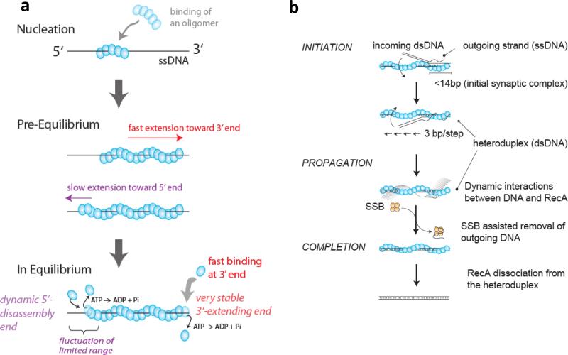
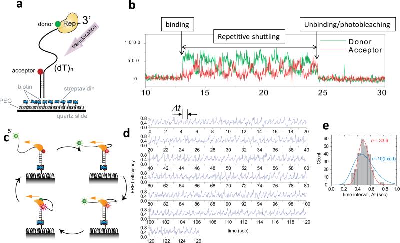
The advent of new technologies allowing the study of single biological molecules continues to have a major impact on studies of interacting systems as well as enzyme reactions. These approaches (fluorescence, optical, and magnetic tweezers), in combination with ensemble methods, have been particularly useful for mechanistic studies of protein-nucleic acid interactions and enzymes that function on nucleic acids. We review progress in the use of single-molecule methods to observe and perturb the activities of proteins and enzymes that function on flexible single-stranded DNA. These include single-stranded DNA binding proteins, recombinases (RecA/Rad51), and helicases/translocases that operate as motor proteins and play central roles in genome maintenance. We emphasize methods that have been used to detect and study the movement of these proteins (both ATP-dependent directional and random movement) along the single-stranded DNA and the mechanistic and functional information that can result from detailed analysis of such movement.


Figures

Figure 1
Figure 1. Proteins on ssDNA
ssDNA is depicted as a black line. (Center) tangential vectors (t1 and t2) along the contour of ssDNA are correlated if the two points are closer than the persistence length (1-3 nm for ssDNA). (Top) RecA or Rad51 forms a right-handed helical filament on ssDNA. ATP binds to the interface between two adjacent proteins. RecA/Rad51 filaments are negatively regulated by some helicases and can efficiently remove SSB proteins bound to ssDNA during their extension (gray arrows). (Bottom right) A helicase can use ATP hydrolysis to translocate directionally on ssDNA. (Bottom left) SSB/RPA binds and protects ssDNA. For E. coli SSB, ssDNA wraps around the tetrameric protein. SSB can stimulate RecA filament formation by removing DNA secondary structures. The consequence of an encounter between a translocating helicase and an SSB protein is an open question.
Figure 2
Figure 2. Structure and dynamics of E. coli SSB/ssDNA complexes
(a) Model of an SSB tetramer bound to 65 nt of ssDNA (SSB)65 mode) (red) based on a crystal structure(104). (b) Cartoon depiction of an SSB tetramer wrapped by ~ 65 nt of ssDNA. (c) SSB has four C-terminal extensions that bind other proteins. (d) smFRET scheme for monitoring SSB binding to a 70 nt ssDNA. When a single SSB tetramer binds in its (SSB)65 mode, the donor and the acceptor are in close proximity, yielding high FRET. When an additional SSB tetramer binds to the same ssDNA with both tetramers in the (SSB)35 mode, a lower FRET value results. The DNA is tethered to a polymer-passivated surface, and extensive comparison to bulk solution data showed that fluorescence labeling and surface tethering did not perturb the reaction. (e) smFRET time traces showing switching between (SSB)65 and (SSB)35 modes induced by dissociation and binding of a second SSB tetramer (Figures adapted from Ref. (113).
Figure 3
Figure 3. smFRET fluctuations arising from diffusional migration of E. coli SSB on ssDNA
(a) –(Top) Rapid fluctuations between several FRET states are observed due to diffusion of an SSB tetramer on an 81 nucleotide ssDNA; (bottom) when the available ssDNA is limited to 69 nucleotides by forming a duplex with the 12 nucleotide extension, the FRET fluctuations are severely reduced reflecting less protein diffusion; (b) – anti-correlated FRET time traces from a three color experiment showing SSB tetramer diffusion (Donor (Alexa555)-labeled SSB bound to a (dT)130 DNA labeled at each end with acceptors (Cy5 and Cy5.5) Figures adapted from Ref. (114).
Figure 4
Figure 4. smFRET studies support a sliding model for SSB diffusion on ssDNA
(a) -Rolling mechanism for SSB diffusion. One end of the wrapped ssDNA could partially dissociate from the SSB resulting in an available site on the SSB to which the free ssDNA at the other end can bind. There would only be relative movement between the donor (green) on the SSB surface and the acceptor (red) on the ssDNA if the acceptor is near the DNA end, but not the midsection. (b) - Sliding mechanism for SSB diffusion. The entire ssDNA (65 nt) moves relative to the SSB protein surface during sliding, hence relative movement between the donor and acceptor should occur for all acceptor positions on the DNA. (c) and (d) - Representative single-molecule time traces of donor(Alexa555) and acceptor(Cy5) intensities and the corresponding FRET efficiencies show the same level of FRET fluctuations regardless of the acceptor (Cy5) position on the DNA. Figures adapted from Ref. (148).
Figure 5
Figure 5. A combined smFRET-force experiment supports a reptation mechanism for SSB sliding on ssDNA
(a) - SSB diffusion persists even with ssDNA under tension. SSB diffusion-induced FRET fluctuations continue for forces up to 5 pN. Magenta arrows indicate SSBf dissociation events. (b) - a reptation (sliding-with-bulge) mechanism for SSB diffusion is proposed to occur by nucleating a thermally activated bulge that propagates via a random walk around the SSB surface. The bulge diffuses back and forth over the entire SSB surface, and if it emerges on the other side, the SSB moves along the ssDNA by one step (~3 nucleotides). The arrows represent the DNA movements and the cyan asterisk represents a single nucleotide position on the DNA. The asterisk-marked position on the ssDNA will move relative to the protein surface. Figures adapted from Ref. (148).
Figure 6
Figure 6. Dynamics of RecA filament formation and strand exchange reactions
(a) RecA filament formations on ssDNA is nucleated by the binding of a RecA oligomer, likely between 4-5 monomers(37; 59), and is extended rapidly in monomer un units mainly in the 3’ direction with a 10 times slower extension in the 5’ direction(59). In the steady state, RecA monomers can dissociate from both ends as monomers upon ATP hydrolysis(59). (b) RecA-ssDNA filament binds to a homologous dsDNA forming an initial synaptic complex (< 14 bp in length), then propagates rapidly in 3 bp increments while undergoing base pair exchange. The outgoing ssDNA displays dynamic interactions with the resulting RecA-dsDNA filament and its removal is facilitated by SSB. Finally, RecA dissociates from the heteroduplex product, completing the reaction (Adapted from Ref. (105)).
Figure 7
Figure 7. Repetitive ssDNA translocation of Rep and PcrA
(a) A donor-labeled Rep monomer translocates on an acceptor-labeled DNA in the 3’ to 5’ direction. (b) Initial binding of Rep is detected as a sudden increase in fluorescence. This is followed by a gradual increase in the acceptor intensity (red) and a concomitant decrease in the donor intensity (green) due to Rep translocation on ssDNA. The protein then snaps back to the initial low FRET state and repeats the cycle until protein unbinding or photobleaching of the donor terminates the fluorescence signal. (c) A cartoon showing how a PcrA monomer can anchor itself to a ds/ssDNA junction and use its ssDNA translocation activity to reel in a 5’ ssDNA tail. Once it runs off the tail end it can re-initiate the repetitive looping cycle from the junction. (d) Representative smFRET time traces of repetitive ssDNA looping (256 cycles) induced by a single molecule of PcrA. Δt denotes the time interval of each cycle. (e) Δt histogram from a single PcrA molecule showing 256 cycles of looping. A fit to the Gamma distribution gives the number of hidden steps, n=33.6, for 40 nt ssDNA (red), supporting a single nt kinetic step size. A forced fit with a fixed n value of 10 to mimic a 4 nt kinetic step size gives a much poorer fit (blue). Figures were adapted from Refs (92; 100).

References

    1. Adzuma K. No sliding during homology search by RecA protein. J Biol Chem. 1998;273:31565–73. - PubMed
    1. Ali JA, Lohman TM. Kinetic Measurement of the Step Size of DNA Unwinding By Escherichia Coli Uvrd Helicase. Science. 1997;275:377–80. - PubMed
    1. Anand SP, Zheng H, Bianco PR, Leuba SH, Khan SA. DNA helicase activity of PcrA is not required for the displacement of RecA protein from DNA or inhibition of RecA-mediated strand exchange. J Bacteriol. 2007;189:4502–9. - PMC - PubMed
    1. Antony E, Tomko EJ, Xiao Q, Krejci L, Lohman TM, Ellenberger T. Srs2 disassembles Rad51 filaments by a protein-protein interaction triggering ATP turnover and dissociation of Rad51 from DNA. Mol Cell. 2009;35:105–15. - PMC - PubMed
    1. Arata H, Dupont A, Mine-Hattab J, Disseau L, Renodon-Corniere A, et al. Direct observation of twisting steps during Rad51 polymerization on DNA. Proc Natl Acad Sci U S A. 2009;106:19239–44. - PMC - PubMed

Publication types

MeSH terms