. 2012 Mar 9;4(1):3.
doi: 10.1186/2045-824X-4-3.

Specific tumor-stroma interactions of EBV-positive Burkitt's lymphoma cells in the chick chorioallantoic membrane

Affiliations

Specific tumor-stroma interactions of EBV-positive Burkitt's lymphoma cells in the chick chorioallantoic membrane

Jürgen Becker et al. Vasc Cell. .

Abstract

Background: Burkitt's lymphoma (BL) is an aggressive Non-Hodgkin lymphoma. Epstein-Barr Virus (EBV) is able to transform B cells and is a causative infectious agent in BL. The precise role of EBV in lymphoma progression is still unclear. Most investigations have concentrated on cell autonomous functions of EBV in B cells. Functions of the local environment in BL progression have rarely been studied, mainly due to the lack of appropriate in vivo models. Therefore, we inoculated different human BL cell-lines onto the chorioallantoic membrane (CAM) of embryonic day 10 (ED10) chick embryos and re-incubated until ED14 and ED17.

Results: All cell-lines formed solid tumors. However, we observed strong differences in the behavior of EBV+ and EBV- cell-lines. Tumor borders of EBV+ cells were very fuzzy and numerous cells migrated into the CAM. In EBV- tumors, the borders were much better defined. In contrast to EBV- cells, the EBV+ cells induced massive immigration of chick leukocytes at the tumor borders and the development of granulation tissue with large numbers of blood vessels and lymphatics, although the expression of pro- and anti-angiogenic forms of Vascular Endothelial Growth Factors/receptors was the same in all BL cell-lines tested. The EBV+ cell-lines massively disseminated via the lymphatics and completely occluded them.

Conclusions: Our data suggest that the EBV+ cells attract pro-angiogenic leukocytes, which then induce secondary tumor-stroma interactions contributing to the progression of BL. We show that the CAM is a highly suitable in vivo model to study the differential behavior of BL cell-lines.


Figures

Figure 1
Figure 1
BL2 cells stained with cell-tracker (green) and incubated on the CAM for 4 days. A) The cells form a solid tumor with almost sharp borders. CAM vessels are faintly visible. Bar = 2 mm. B) Higher magnification of A) showing a few green tumor cells at some distance from the solid tumor. Bar = 150 μm.
Figure 2
Figure 2
Staining of metastatic BL2 cells in the CAM. A) BL2 cells are marked with cell-tracker green. B) BL2 cells stain with anti-human-HLA antibodies (red). C) Staining of all nuclei with Dapi (blue). D) Merged picture. Bar = 60 μm.
Figure 3
Figure 3
BL2B95 cells stained with cell-tracker (green) and incubated on the CAM for 4 days. The cells form solid tumors. The tumor borders are less sharp than those of BL2 cells. CAM vessels are faintly visible. A) A great mass of tumor cells (asterisk) is seen adjacent to the solid tumor (T). Bar = 2,5 mm B) Higher magnification of A). Note tumor cells (arrows) that obviously migrate from the tumor. Bar = 150 μm. C) Numerous scattered lymphoma cells and clusters of lymphoma cells are present at some distance from the solid tumor (T). Bar = 300 μm. D) Note lymphoma cells (arrows) lining up along a CAM vessel. Bar = 150 μm.
Figure 4
Figure 4
BL2B95 cells stained with cell-tracker (green) form solid tumors (T) on the CAM after 4 days. Clusters of lymphoma cells (arrows) have invaded the CAM. Mep 21 staining (red) reveals a large amount of blood capillaries in the CAM. Bar = 100 μm.
Figure 5
Figure 5
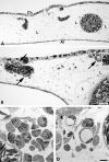
Semi- and ultrathin sections of CAM tissue approximately 7 mm apart from the solid tumor. A) In BL2 specimens the typical morphology of the CAM is found with capillaries located in the chorionic epithelium (Ch), larger blood vessels and lymphatics (L) in the stroma, and the allantoic epithelium (Al). Bar = 80 μm. B) In BL2B95 specimens large numbers of leukocytes (arrows) are found in perivascular position. C, D) Ultra-thin sections of the specimen shown in B). Note invasion of granulocytes and other leukocytes into the CAM stroma. Bar = 10 μm.
Figure 6
Figure 6
Semi-thin sections of BL2 tumors. A) Tumor cells were applied on the CAM within Matrigel, which is still visible after 7 days as amorphous material (M). Ch, chorionic epithelium; Al, allantoic epithelium. Insert: Higher magnification of lymphoma cells in Matrigel. Note the large nuclei with one or several nucleoli. Bar = 120 μm. B) Higher magnification of A) showing tumor cells in Matrigel (M). Tumor cells have invaded the CAM stroma (St). Blood vessels (arrows); Mitotic figures of lymphoma cells (arrowheads). Bar = 30 μm.
Figure 7
Figure 7
Semi-thin sections of BL74 and BL2B95 tumors. A) BL74 cells were applied on the CAM within Matrigel (M), which is visible after 4 days as amorphous material. Al, allantoic epithelium. Note the granulation tissue in the CAM stroma (St). Bar = 120 μm B) Higher magnification of A) showing tumor cells in Matrigel (M). Note the large number of leukocytes (small round cells) and the numerous blood vessels (arrows) in the CAM stroma. C) Peritumoral stroma of a BL2B95 tumor. Note large numbers of blood vessels and lymphatics (L). Bar = 30 μm in B,C. D) Ultra-thin section showing tumor cells (T) in the CAM stroma immediately accompanied by chick leukocytes (arrowheads). Bar = 15 μm.
Figure 8
Figure 8
Semi- and ultra-thin sections of BL2B95 tumors. A) At approximately 5 mm distance from the solid tumor, some lymphoma cells are found in the CAM stroma. The CAM lymphatics (L) are completely filled with tumor cells. Bar = 300 μm. B - D) Higher magnification of the specimen in A). B, C) Semi-thin sections. The lymphatics (L) are completely filled with tumor cells. Bar = 30 μm in B, and 40 μm in C. D) Ultra-thin section showing lymphatic endothelium (LE) and tumor cells, which fill the vessel completely. Two leukocytes (arrowheads) are visible. One is in contact with a dying lymphoma cell (asterisk), as seen by the heterochromatin condensation in the periphery of the nucleus. Bar = 10 μm.
Figure 9
Figure 9
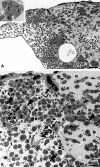
Semi-thin sections of BL2 tumors. A) Immediately adjacent to the solid tumor (on the left side of the specimen), numerous lymphoma cells have invaded the CAM stroma. Only very few cells are found in the CAM lymphatics (L). Bar = 400 μm. B) Higher magnification of A) showing CAM lymphatics (L) invaded by only a few tumor cells (arrows). Bar = 80 μm.
Figure 10
Figure 10
Real-time RT-PCR of BL2 and BL2B95 lymphoma cells in comparison with neuroblastoma (NB) cells. Expression of A) VEGF-A, B) VEGF-C, C) VEGF-D, D) VEGFR-1, E) sVEGFR-1 and F) esVEGFR-2. As compared to NB cells Shep and SK-IN there is no significant expression of the growth factors in the BL2 and BL2B95 cells. Membrane-bound and soluble forms of VEGFR-1 are weakly and equally expressed in the lymphoma cell-lines, while esVEGFR-2 is not expressed.

Similar articles

Cited by

References

    1. Pfreundschuh M, Schubert J, Ziepert M, Schmits R, Mohren M, Lengfelder E, Reiser M, Nickenig C, Clemens M, Peter N. et al.Six versus eight cycles of bi-weekly CHOP-14 with or without rituximab in elderly patients with aggressive CD20+ B-cell lymphomas: a randomised controlled trial (RICOVER-60) Lancet Oncol. 2008;9:105–116. doi: 10.1016/S1470-2045(08)70002-0. - DOI - PubMed
    1. Kaiser U, Uebelacker I, Abel U, Birkmann J, Trumper L, Schmalenberg H, Karakas T, Metzner B, Hossfeld DK, Bischoff HG. et al.Randomized study to evaluate the use of high-dose therapy as part of primary treatment for "aggressive" lymphoma. J Clin Oncol. 2002;20:4413–4419. doi: 10.1200/JCO.2002.07.075. - DOI - PubMed
    1. Wunderlich A, Kloess M, Reiser M, Rudolph C, Truemper L, Bittner S, Schmalenberg H, Schmits R, Pfreundschuh M, Loeffler M. Practicability and acute haematological toxicity of 2- and 3-weekly CHOP and CHOEP chemotherapy for aggressive non-Hodgkin's lymphoma: results from the NHL-B trial of the German High-Grade Non-Hodgkin's Lymphoma Study Group (DSHNHL) Ann Oncol. 2003;14:881–893. doi: 10.1093/annonc/mdg249. - DOI - PubMed
    1. Burkitt D. A Sarcoma Involving the jaws in African Children. Br J Surg. 1958;46:218–223. doi: 10.1002/bjs.18004619704. - DOI - PubMed
    1. Chene A, Donati D, Orem J, Mbidde ER, Kironde F, Wahlgren M, Bejarano MT. Endemic Burkitt's lymphoma as a polymicrobial disease: new insights on the interaction between Plasmodium falciparum and Epstein-Barr virus. Semin Cancer Biol. 2009;19:411–420. doi: 10.1016/j.semcancer.2009.10.002. - DOI - PubMed

LinkOut - more resources