. 2012 Jun;11(3):542-52.
doi: 10.1111/j.1474-9726.2012.00816.x. Epub 2012 Apr 4.

Aging brain microenvironment decreases hippocampal neurogenesis through Wnt-mediated survivin signaling

Affiliations

Aging brain microenvironment decreases hippocampal neurogenesis through Wnt-mediated survivin signaling

Carlos J Miranda et al. Aging Cell. 2012 Jun.

Abstract

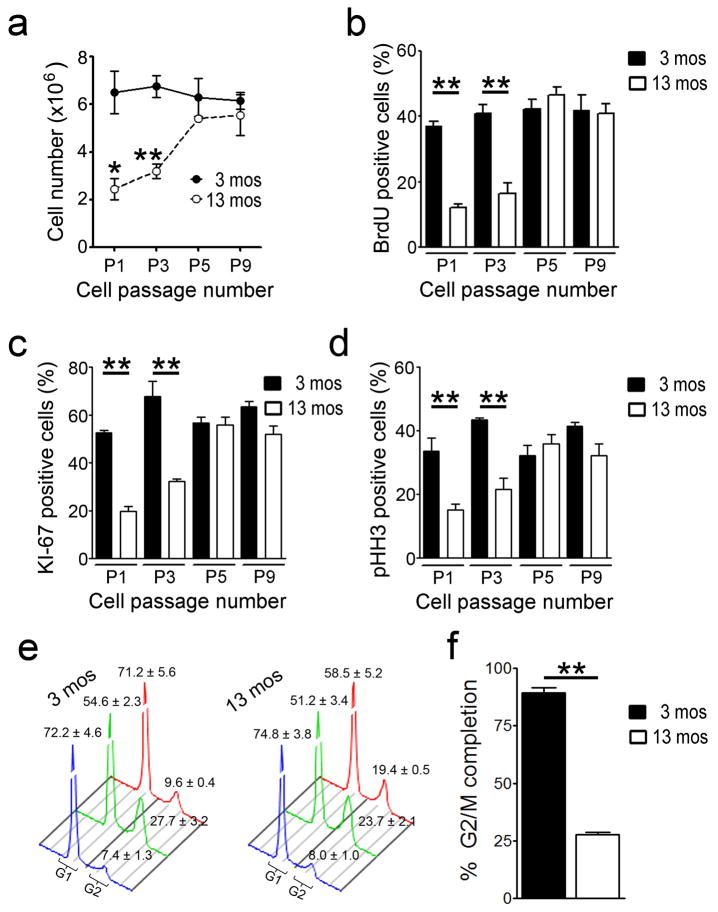
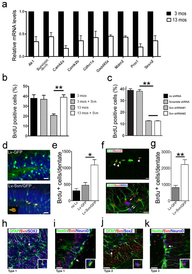
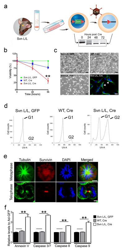
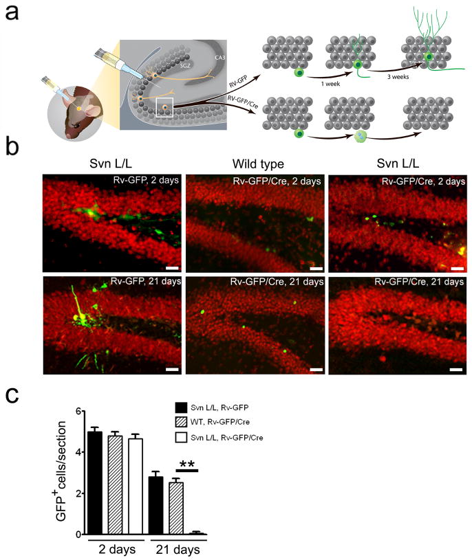
Accumulating evidence suggests that adult hippocampal neurogenesis relies on the controlled and continued proliferation of neural progenitor cells (NPCs). With age, neurogenesis decreases through mechanisms that remain unclear but are believed to involve changes in the NPC microenvironment. Here, we provide evidence that NPC proliferation in the adult brain is in part regulated by astrocytes via Wnt signaling and that this cellular cross-talk is modified in the aging brain, leading to decreased proliferation of NPCs. Furthermore, we show that astrocytes regulate the NPC cell cycle by acting on the expression levels of survivin, a known mitotic regulator. Among cell cycle genes found down-regulated in aged NPCs, survivin was the only one that restored NPC proliferation in the aged brain. Our results provide a mechanism for the gradual loss of neurogenesis in the brain associated with aging and suggest that targeted modulation of survivin expression directly or through Wnt signaling could be used to stimulate adult neurogenesis.


Figures

Fig. 1
Fig. 1
Proliferation deficit in aged NPCs is reversed with time in culture. (a) Total number of cells from 3- and 13-month NPCs cultures with time in culture. At each cell passage (P), cells were counted and replated at 1×10⁶ cells/10cm dish. Cells were passaged every 4 days. (b–d) Levels of cell proliferation observed in 3- and 13-month NPCs with time in culture as determined by the percentage of cells positive for the S-phase mitotic marker BrdU (b), the pan-cell cycle marker KI-67 (c), or the G2/M cell cycle marker pHH3 (d). (e) Cell cycle profiles of 3- and 13 month NPCs under proliferative conditions (blue), following nocodazole treatment (green), and after release from nocodazole block (red). (f) Percentage of cells that have completed the G2/M phase of the cell cycle as show in (e). Mean values ± SEM from triplicate experiments are shown; * P < 0.05; ** P< 0.01.
Fig. 2
Fig. 2
Survivin levels in NPCs are decreased with aging and regulate proliferation. (a) Cell cycle regulatory genes down-regulated in 13-month compared to 3-month NPCs as determined by qRT-PCR. Gene analysis was performed P2. (b) Effects of exogenous survivin expression on the proliferation of 3- and 13-month NPCs (P3). (c) Effects of survivin knock-down on the proliferation of 3-month-NPCs. (d) Representative image showing transduction of cells with lentivirus expressing GFP (Lv-GFP) or survivin-GFP (Lv-Svn/GFP) in the granular layer and SGZ. BrdU+ cells were found only in the SGZ and co-localized with GFP- expressing cells. (e) Quantification of BrdU+ cells in the DG of control, Lv-GFP injected, or Lv-Svn/GFP injected 13-month mice during a daily seven day BrdU pulse. N = 6 mice per group. (f) Identity of BrdU+ cells transduced with Lv-Svn/GFP. Type 1 (double arrow), Type 2 (single arrow) and Type 3 (arrowheads) NPCs are indicated. (g) Quantification of BrdU+ cells in the DG of Lv-GFP or Lv-Svn/GFP injected 13-month mice during a 24 hours BrdU pulse. Survivin expression can be found in Sox2+, GFAP+, Nestin+, NeuroD- Type 1 NPCs (h–i) Sox2+, GFAP- Type 2 NPCs (j) Nestin-, NeuroD+ Type 3 NPCs (k). Cells identified by the arrows in panels h–k are shown magnified in the accompanied figure inset. In all graphs, mean ± SEM from triplicates are shown, *P < 0.05. ** P < 0.01. Scale bars, 20 μm.
Fig. 3
Fig. 3
Survivin is required for the proliferation and survival of NPCs. (a) A schematic of the experimental procedure used to ablate survivin in NPCs. Survivin was deleted using adenoviral mediated expression of Cre-recombinase. (b) Cell viability was determined by the MTT assay in survivin expressing NPCs (svnL/L – GFP, Green and wild type – Cre, Blue) and survivin-deficient NPCs (svnL/L -Cre, Red). (c) Phase contrast and fluorescence images showing decreased cell viability and presence of cells with two nuclei in survivin-deficient NPCs (bottom panel) isolated from svnL/L mice 72 hours post Ad-Cre infection compared with Ad-GFP in svnL/L or Ad-Cre in wild-type NPCs (upper panel). (d) Cell cycle profiles as obtained by flow cytometry analysis of DNA content of proliferating svnL/L NPCs treated with Cre showing arrest in G2/M compared to wild-type treated controls. (e) Intracellular localization of endogenous survivin in NPCs during mitosis. Scale bars, 5 μm. (f) Levels of annexin V positive cells and caspase activation found in svnL/L NPCs culture 72 hours post Cre induction. In all graphs, mean ± SEM from triplicates is shown, ** P< 0.01.
Fig. 4
Fig. 4
Creation of newborn adult hippocampal neurons requires survivin expression in NPCs. (a) Schematic of the experimental approach depicting dividing NPCs in the SGZ transduced with retroviruses (Rv) carrying GFP (cytoplasmic localization) or a Cre-GFP fusion protein (nuclear localization). Hippocampi were analyzed 3 weeks later for the presence of GFP+ granular neurons recognized by NeuN expression. (b) Similar numbers of NPCs were transduced with Rv-GFP or Rv-GFP/Cre and expressed GFP two days post-injection into wild type or svnL/L mice (upper panels). After 3 weeks (bottom panels), approximately 50% of cells initially transduced with Rv-GFP in svnL/L mice or with Rv-GFP/Cre in wild type mice were still present in the granular layer and express NeuN. By contrast, in Rv-GFP/Cre injected svnL/L mice no GFP-transduced cells were observed at 3 weeks (bottom middle panel). (c) Quantification of GFP positive cells found following Rv-GFP and Rv-GFP/Cre at day 2 and 21 as in “b”. ** P<0.001, n= 8 for each time point). Mean ± SEM are shown.
Fig. 5
Fig. 5
The neurogenic microenvironment of older mice represses NPC survivin expression and NPC proliferation. (a) Survivin promoter activity (red) in 3- and 13-month GFP+ NPCs (green) transplanted into the DG of 3- or 13-month old mouse brains. Representative images (n = 6 mice) are shown for each experimental group. (b) Quantification of red fluorescence levels in transplanted NPC cells visualized in “a”. Bottom numbers indicate age in months of transplanted NPCs and recipient mouse DG. (c–d) Effect of conditioned medium (CM) from 3- or 13- month astrocytes on survivin promoter activity (c; firefly luciferase reporter activity) and proliferation (d; BrdU incorporation) in 3- and 13-month NPCs. Mean percentages ± SEM from triplicate experiments are shown. * P < 0.05; ** P < 0.01. Scale bars, 20 μm.
Fig. 6
Fig. 6
Age dependent reduction of astrocyte mediated Wnt signaling correlates with a decrease in survivin levels and proliferation of NPCs. (a) Quantitative RT-PCR comparison of Wnt family gene expression levels between 3- and 13-month astrocytes (Ast). (b–c) Effects of Wnts and Wnt inhibitors (sFRPs) on survivin promoter activity (b) and proliferation (c) in 3- month NPCs in the presence of conditioned medium (CM) from 3- or 13-month astrocytes (Ast). Growth medium (GM) contains FGF2 and EGF. (d) Survivin knockdown abolishes Wnt effect in augmenting NPC proliferation. Mean values ± SEM from triplicates are shown. ** P < 0.01.

Similar articles

Cited by

References

    1. Alvarez-Buylla A, Temple S. Stem cells in the developing and adult nervous system. J Neurobiol. 1998;36:105–110. - PubMed
    1. Bonaguidi MA, Wheeler MA, Shapiro JS, Stadel RP, Sun GJ, Ming GL, Song H. In vivo clonal analysis reveals self-renewing and multipotent adult neural stem cell characteristics. Cell. 2011;145:1142–1155. - PMC - PubMed
    1. Caldas H, Jiang Y, Holloway MP, Fangusaro J, Mahotka C, Conway EM, Altura RA. Survivin splice variants regulate the balance between proliferation and cell death. Oncogene. 2005;24:1994–2007. - PubMed
    1. Encinas JM, Michurina TV, Peunova N, Park JH, Tordo J, Peterson DA, Fishell G, Koulakov A, Enikolopov G. Division-coupled astrocytic differentiation and age-related depletion of neural stem cells in the adult hippocampus. Cell Stem Cell. 2011;8:566–579. - PMC - PubMed
    1. Esteve PO, Chin HG, Pradhan S. Human maintenance DNA (cytosine-5)-methyltransferase and p53 modulate expression of p53-repressed promoters. Proc Natl Acad Sci U S A. 2005;102:1000–1005. - PMC - PubMed

Publication types