. 2012 Aug;11(4):569-78.
doi: 10.1111/j.1474-9726.2012.00818.x. Epub 2012 Apr 17.

Glucocorticoids suppress selected components of the senescence-associated secretory phenotype

Affiliations

Glucocorticoids suppress selected components of the senescence-associated secretory phenotype

Remi-Martin Laberge et al. Aging Cell. 2012 Aug.

Abstract

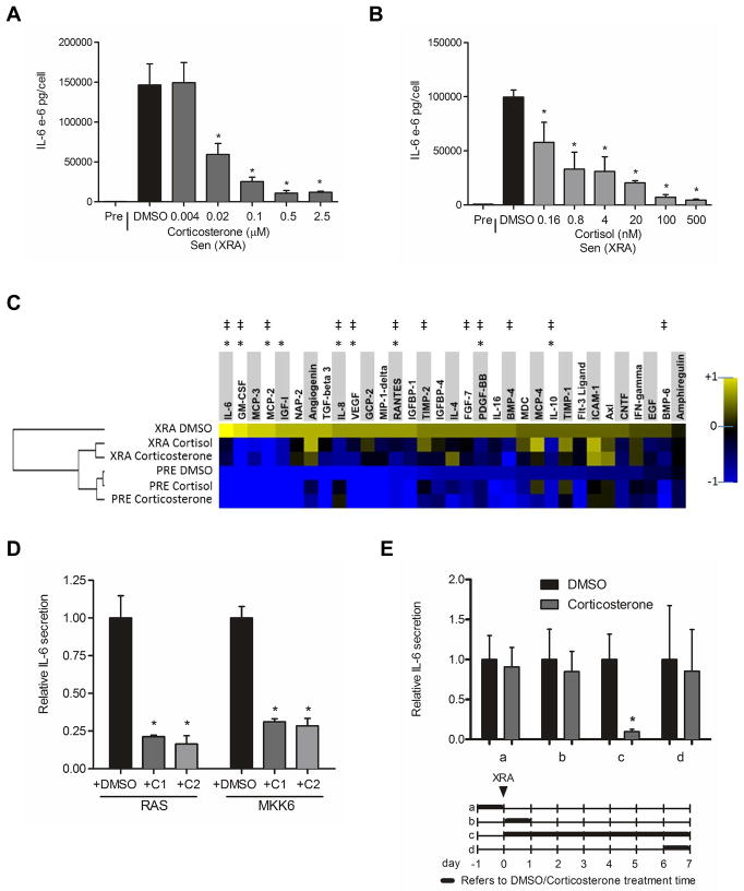
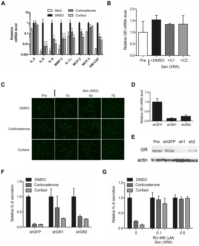
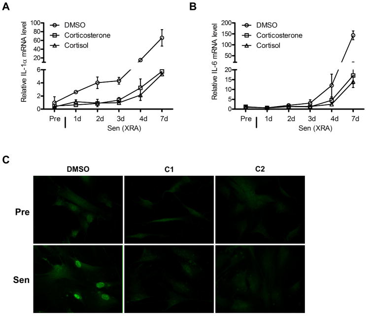
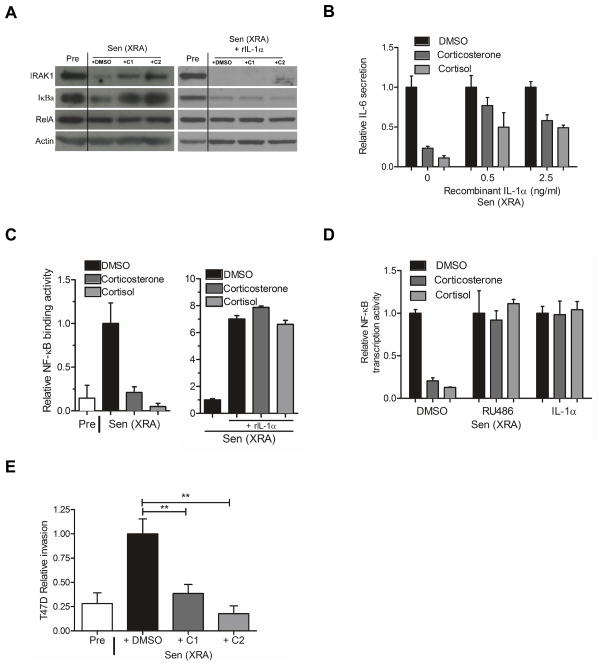
Cellular senescence suppresses cancer by arresting the proliferation of cells at risk for malignant transformation. Recently, senescent cells were shown to secrete numerous cytokines, growth factors, and proteases that can alter the tissue microenvironment and may promote age-related pathology. To identify small molecules that suppress the senescence-associated secretory phenotype (SASP), we developed a screening protocol using normal human fibroblasts and a library of compounds that are approved for human use. Among the promising library constituents was the glucocorticoid corticosterone. Both corticosterone and the related glucocorticoid cortisol decreased the production and secretion of selected SASP components, including several pro-inflammatory cytokines. Importantly, the glucocorticoids suppressed the SASP without reverting the tumor suppressive growth arrest and were efficacious whether cells were induced to senesce by ionizing radiation or strong mitogenic signals delivered by oncogenic RAS or MAP kinase kinase 6 overexpression. Suppression of the prototypical SASP component IL-6 required the glucocorticoid receptor, which, in the presence of ligand, inhibited IL-1α signaling and NF-κB transactivation activity. Accordingly, co-treatments combining glucocorticoids with the glucocorticoid antagonist RU-486 or recombinant IL-1α efficiently reestablished NF-κB transcriptional activity and IL-6 secretion. Our findings demonstrate feasibility of screening for compounds that inhibit the effects of senescent cells. They further show that glucocorticoids inhibit selected components of the SASP and suggest that corticosterone and cortisol, two FDA-approved drugs, might exert their effects in part by suppressing senescence-associated inflammation.


Conflict of interest statement

The authors report no financial or other conflict of interest relevant to the subject of this article.

Figures

Figure 1
Figure 1. Corticosterone and cortisol partially suppress the SASP
(A) Senescent (X-irradiated with 10 Gy, (Sen (XRA)) HCA2 fibroblasts were incubated in medium plus 10% serum containing the indicated concentrations of corticosterone or the highest concentration of DMSO (vehicle control). The cells were given corticosterone or DMSO immediately after irradiation and analyzed 6 d later. The cells were washed and incubated in serum-free medium without corticosterone to generate conditioned media. Conditioned media from presenescent (Pre) and control or corticosterone-treated Sen (XRA) cells were analyzed by ELISA for IL-6. (B) Cells were treated and conditioned media were generated and analyzed as described in (A) except cortisol was used at the indicated concentrations. (C) Conditioned media were collected from presenescent (PRE) or senescent (XRA) cells that were treated with DMSO, corticosterone (500 nM) or cortisol (100 nM) as described in (A). The conditioned media were analyzed by antibody arrays. We used the average signal from PRE and XRA DMSO cells as the baseline. Signals higher than baseline are yellow; signals lower than baseline are blue. Color intensities represent log2-fold changes from the average value. The hierarchical clustering relationship between samples is shown as a dendrogram (left). *, factors significantly (p<0.05) suppressed by cortisol. ‡, factors significantly (p<0.05) suppressed by corticosterone. (D) Cells were infected with RAS- or MKK6-expressing lentiviruses. After selection, the cells were given DMSO-, 500 nM corticosterone (C1)- and 100 nM cortisol (C2) for 6 d. Conditioned media were generated as described above and analyzed by ELISA for IL-6. *, factors significantly different from DMSO-treated (p<0.05). (E) Cells were treated with 500 nM corticosterone for the indicated intervals (a–d, indicated by the thick lines in the lower panel) before or after X-irradiation (XRA, indicated by the arrow). Conditioned media were prepared and analyzed by ELISA for IL-6 (upper panel). *, factors significantly different from DMSO-treated (p<0.05).
Figure 2
Figure 2. Effect of glucocorticoids on the SASP depends on the glucocorticoid receptor
(A) mRNA was extracted from presenescent (Mock) or senescent X-irradiated HCA2 cells treated with DMSO, 500 nM corticosterone or 100 nM cortisol as described in the legend to Figure 1. Transcripts for IL-5, IL-6, IL-8, MMP-3, IL-1α, MCP-2, MCP-3 and GM-CSF were quantified by quantitative PCR (normalized to tubulin). *, factors significantly different from DMSO-treated (p<0.05). (B) mRNA was extracted from Pre and Sen (XRA) cells treated with DMSO, 500 nM corticosterone (C1) or 100 nM cortisol (C2) as described above, and transcripts for the GR were quantified by PCR (normalized to tubulin). Although GR mRNA levels tended to be slightly elevated in senescent cells, the increase was not statistically significant. (C) Pre and Sen (XRA) cells treated with DMSO, 500 nM corticosterone or 100 nM cortisol as described above were immunostained for GR 1, 4 and 7 d after X-irradiation. (D) Cells were infected with lentiviruses expressing shRNAs against GFP (control) or GR, and selected. Seven days after selection, mRNA was extracted and transcripts for GR were quantified by PCR (normalized to tubulin). (E) Total cell lysates were prepared from the shGFP- and shGR-expressing cells described in (D), and analyzed by western blotting for GR and actin (control). (F) Cells infected with shGFP or shGR-expressing lentiviruses were X-irradiated and treated immediately thereafter with DMSO, 500 nM corticosterone or 100 nM cortisol. Conditioned media were collected 7 d later and analyzed by ELISA for IL-6. (G) Cells were treated as described in (F) except for the addition of RU-486 at the indicated doses. Conditioned media were collected and analyzed by ELISA for IL-6 secretion.
Figure 3
Figure 3. Glucocorticoids repress IL-1α expression
(A) Presenescent (Pre) HCA2 cells were treated with DMSO, 500 nM corticosterone or 100 nM cortisol for 24 h, or were induced to senesce by X-irradiation (Sen (XRA)) and given DMSO, corticosterone or cortisol immediately thereafter. mRNA was extracted after the indicated intervals and transcripts for IL-1α were quantified by PCR (normalized to tubulin). (B) mRNA extracted from cells described in (A) was used to quantify transcripts for IL-6 (normalized to tubulin). (C) Pre and Sen (XRA) cells, prepared as described in (A), were immunostained for IL-1α. Sen (XRA) cells were immunostained 7 d after irradiation.
Figure 4
Figure 4. Glucocorticoids impair the IL-1α/NF-κB pathway and suppress the ability of the SASP to induce tumor cell invasion
(A) Total HCA2 cell lysates were prepared from presenescent (Pre) cells, or senescent cells (Sen (XRA)) cells treated with DMSO-, 500 nM corticosterone (C1)-, or 100 nM cortisol (C2) in the absence (left panel) or presence (right panel) of recombinant IL-1α protein (rIL-1α). The lysates were analyzed by western blotting for IRAK1, IkBα, RelA and actin (control). (B) After irradiation, Sen (XRA) cells were given DMSO, 500 nM corticosterone or 100 nM cortisol. Six d later, the cells were given recombinant IL-1α protein at the indicated doses in the presence of the glucocorticoids in serum free media. Conditioned media were collected 24 h later and analyzed by ELISA for IL-6. (C) Nuclear extracts were prepared from Pre cells, and Sen (XRA) cells treated with DMSO, 500 nM corticosterone or 100 nM cortisol in the absence (left panel) or presence (right panel) of recombinant IL-1α protein (rIL-1α), and analyzed for NF-κB DNA binding activity. (D) Cells were infected with a lentivirus carrying an NF-κB-luciferase reporter construct, irradiated, and allowed to senesce. Immediately after irradiation, cells were treated with DMSO, 500 nM corticosterone or 100 nM cortisol, plus 0.5 μM RU-486 or 2.5 ng/ml IL-1α, as indicated. Seven d after irradiation, cells were trypsinized, counted, lysed and assayed for luciferase activity, which was normalized to cell number. (E) We prepared conditioned media from presenescent (Pre) cells or senescent cells (Sen (XRA) that had been treated with corticosterone (C1) or cortisol (C2) as described in the legend to Figure 1. The conditioned media were then assayed for ability to stimulate T47D human breast cancer cells to invade a basement membrane, as described in the Experimental Procedures.

References

    1. Acosta JC, O’Loghlen A, Banito A, Guijarro MV, Augert A, Raguz S, Furnagalli M, DaCosta M, Brown C, Popov N, Takastu, Yabuta N, Melamed J, d’Adda di Fagagna F, Bernard D, Hernando E, Gil J. Chemokine signaling via the CXCR2 receptor reinforces senescence. Cell. 2008;133:1006–1018. - PubMed
    1. Adams PD. Healing and hurting: molecular mechanisms, functions and pathologies of cellular senescence. Molec Cell. 2009;36:2–14. - PubMed
    1. Baker DJ, Wijshake T, Tchkonia T, LeBrasseur NK, Childs BG, van de Sluis B, Kirkland JL, van Deursen JM. Clearance of p16Ink4a-positive senescent cells delays ageing-associated disorders. Nature. 2011;479:232–236. - PMC - PubMed
    1. Bartholomew JN, Volonte D, Galbiati F. Caveolin-1 regulates the antagonistic pleiotropic properties of cellular senescence through a novel Mdm2/p53-mediated pathway. Cancer Res. 2009;69:2878–2886. - PMC - PubMed
    1. Beausejour CM, Campisi J. Ageing: balancing regeneration and cancer. Nature. 2006;443:404–405. - PubMed

Publication types

MeSH terms