. 2012 Mar 9:13:7.
doi: 10.1186/1471-2199-13-7.

Protein kinase CK2 localizes to sites of DNA double-strand break regulating the cellular response to DNA damage

Affiliations

Protein kinase CK2 localizes to sites of DNA double-strand break regulating the cellular response to DNA damage

Birgitte B Olsen et al. BMC Mol Biol. .

Abstract

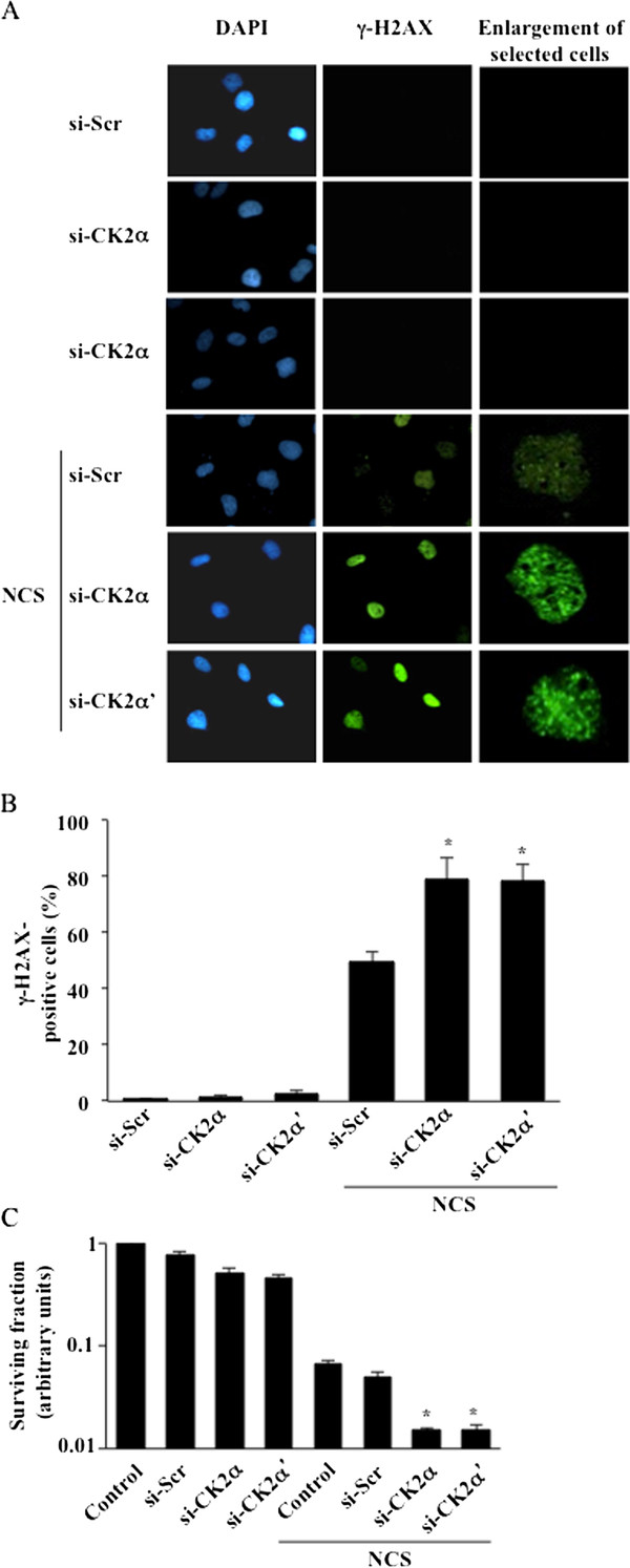
Background: The DNA-dependent protein kinase (DNA-PK) is a nuclear complex composed of a large catalytic subunit (DNA-PKcs) and a heterodimeric DNA-targeting subunit Ku. DNA-PK is a major component of the non-homologous end-joining (NHEJ) repair mechanism, which is activated in the presence of DNA double-strand breaks induced by ionizing radiation, reactive oxygen species and radiomimetic drugs. We have recently reported that down-regulation of protein kinase CK2 by siRNA interference results in enhanced cell death specifically in DNA-PKcs-proficient human glioblastoma cells, and this event is accompanied by decreased autophosphorylation of DNA-PKcs at S2056 and delayed repair of DNA double-strand breaks.

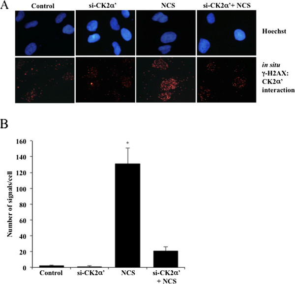
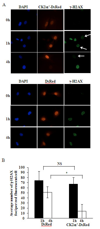
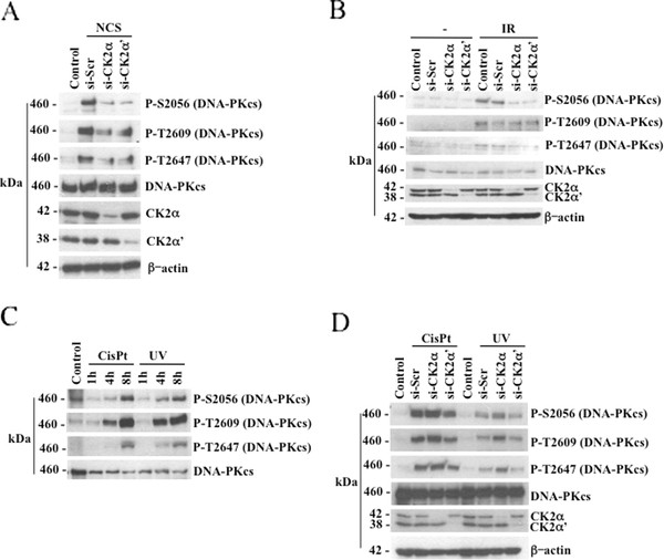
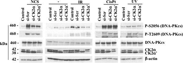
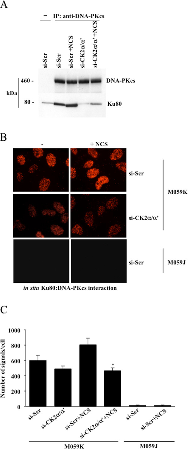
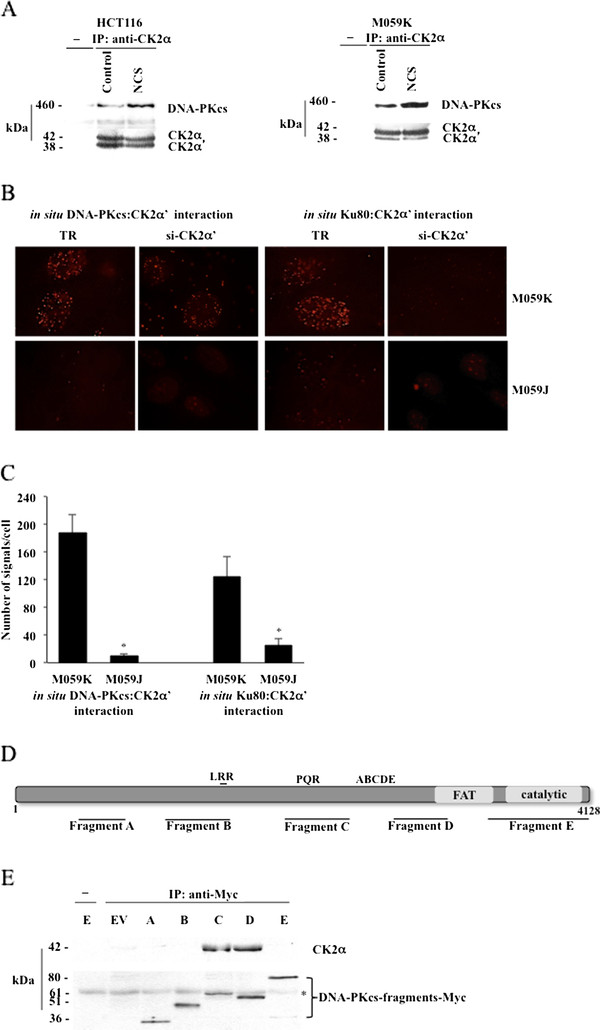
Results: In the present study, we show that CK2 co-localizes with phosphorylated histone H2AX to sites of DNA damage and while CK2 gene knockdown is associated with delayed DNA damage repair, its overexpression accelerates this process. We report for the first time evidence that lack of CK2 destabilizes the interaction of DNA-PKcs with DNA and with Ku80 at sites of genetic lesions. Furthermore, we show that CK2 regulates the phosphorylation levels of DNA-PKcs only in response to direct induction of DNA double-strand breaks.

Conclusions: Taken together, these results strongly indicate that CK2 plays a prominent role in NHEJ by facilitating and/or stabilizing the binding of DNA-PKcs and, possibly other repair proteins, to the DNA ends contributing to efficient DNA damage repair in mammalian cells.


Figures

Figure 1
Figure 1
Down-regulation of CK2 results in persistent γ-H2AX signal and reduced colony formation in human M059K glioblastoma cells. A. Cells were transfected with scramble siRNA (si-Scr), CK2α or -α'-siRNA for 72 hours. Where indicated, 0.5 μg/ml neocarzinostatin (NCS) was added to the medium in the last 24 hours of incubation. Fixed cells were subsequently labeled with anti-γ-H2AX antibody and with a FITC-conjugated secondary antibody. Nuclei were visualized by DAPI staining. B. Quantification of γ-H2AX-positive cells was performed by using ImageJ software and expressed as percentage of the total number of cells in each sample. Bars indicate mean values +/- standard deviation (SD) from three independent experiments. *P < 0.0001 indicates statistically significant difference in the number of γ-H2AX-positive cells in CK2-depleted versus si-Scr-treated cells. C. Cells were transfected with CK2α-, -α'-siRNA or scramble siRNA for 72 hours. 0.5 μg/ml NCS was added in the last hour of incubation. Control, refers to cell treated with transfection reagent only. Cells were allowed to form clusters for 14 days. Colonies were visualized by staining with crystal violet as described in Experimental Procedures. Bar graph shows cell colonies quantification. Average values +/- SD from three independent experiments are shown relative to control (i.e. transfection reagent-treated) cells. *P < 0.005 denotes statistically significant difference in number of colonies formed as compared to si-Scr-tranfected and NCS treated cells.
Figure 2
Figure 2
CK2 co-localizes with γ-H2AX to sites of DNA damage. A. Association between CK2α' and γ-H2AX was revealed by in situ PLA. Cells expressing or lacking CK2α' were left untreated or incubated with 0.5 μg/ml NCS for 1 hour before fixation and labeling with primary antibodies directed against the indicated proteins. Control, refers to cells transfected with si-CK2α' and stained with secondary antibodies. Cell nuclei were revealed by Hoechst staining. B. Quantification of the number of signals/cell as distinct fluorescent red spots was performed by computer-assisted image analysis as described in Experimental Procedures. Mean values +/- SD from three independent experiments are shown. *P < 0.0001 denotes statistically significant difference.
Figure 3
Figure 3
Cells overexpressing CK2 exhibit a prompt response to induction of DNA double-strand break. A. M059K cells transiently overexpressing CK2α'-DsRed or DsRed-fluorescent protein were incubated with 0.5 μg/ml NCS for the indicated times. Subsequently, cells were fixed and labeled with anti-γ-H2AX antibody. Cell nuclei were revealed by DAPI staining. Arrows: cells overexpressing CK2α'-DsRed are more efficient in DNA damage repair as compared to cells that do not overexpress the kinase. One representative experiment is shown. B. Average γ-H2AX foci number per red fluorescent cell as described in A. Mean values +/- SD from three independent experiments are shown. More than 100 cells have been analyzed per experiment. *P < 0.001 denotes statistically significant difference. NS, not significant.
Figure 4
Figure 4
CK2 depletion results in decreased DNA-PKcs phosphorylation in M059K cells following induction of DNA double-strand break. A. Cells were treated as described in Figure 1. Whole cell extracts were employed for Western blot analysis of the indicated proteins. Detection of β-actin was used as loading control. B. Cells were treated essentially as described in Figure 1 except that DNA damage was induced by cell exposure to 10 Gy ionizing radiation. After 1 hour, cells were harvested and total cell lysates were prepared for Western blot analysis. C. M059K cells were treated with 33 μM cisplatin (cisPt) or exposed to 20 J/m2 UV irradiation for the indicated time-points. Cell extract from control and treated cells was prepared and analyzed by Western blot employing antibodies against DNA-PKcs or its phosphorylated forms as indicated in the figure. D. Experiments were performed as described above and in Figure 1 except that cells were treated with cisPt (33 μM, 8 hours incubation) or exposed to UV (20 J/m2, 8 hours recovery) as indicated in the figure. Whole cell lysates were prepared for the analysis by Western blot of DNA-PKcs phosphorylation at S2056, T2609 and T2647. Experiments were performed at least three times obtaining similar results. Data from one representative experiment are shown.
Figure 5
Figure 5
Decreased phosphorylation of DNA-PKcs in CK2-depleted cells is not confined solely to glioblastoma cells. The human non-small cell lung carcinoma H1299 cell line was subjected to treatments described in Figures 1 and 5. Western blot analysis of whole cell lysate was performed employing antibodies against proteins indicated in the figure. Experiments were performed three times obtaining similar results and data from one representative experiment are shown.
Figure 6
Figure 6
siRNA-mediated knock-down of CK2 destabilizes the association between DNA-PKcs and Ku80. A. Cells were transfected with si-Scr and si-CK2α/α', respectively. After 71 hours, cells were incubated with 0.5 μg/ml NCS for 1 additional hour as indicated in the figure. Whole cell lysate was subjected to co-immunoprecipitation (IP) with a rabbit polyclonal anti-DNA-PKcs antibody. Immunoprecipitated proteins were revealed by Western blot employing mouse monoclonal antibodies against the indicated proteins. A control experiment was performed where crude extract from cells transfected with scramble-siRNA was subjected to immunoprecipitation with normal rabbit serum (-). B. In situ association between DNA-PKcs and Ku80 was investigated in M059K- and DNA-PKcs-deficient M059J cells, treated according to conditions indicated in the figure, by in situ PLA. The molecular interaction is indicated by the presence of distinct red fluorescent spots in the cell nuclei. C. Quantification of the number of positive signals/cell was performed by computer-assisted image analysis. Mean values +/- SD from three independent experiments are shown. *P < 0.0001 denotes statistically significant difference between cell populations incubated with 0.5 μg/ml NCS for 1 hour and treated with si-Scr or siRNAs against CK2α and -α', respectively.
Figure 7
Figure 7
DNA-PKcs associates with CK2. The complex formation is enhanced upon induction of DNA damage. A. Cell lysates from HCT116 and M059K cells were employed in co-immunoprecipitation experiments in the presence of normal rabbit serum (-) or rabbit polyclonal anti-CK2α antibody. Immunoprecipitates were analyzed by Western blot with antibodies against the indicated proteins. B. The association between endogenous CK2α' and DNA-PKcs or Ku80 were explored by in situ PLA in M059K cells treated with 0.5 μg/ml NCS for 1 hour. Control experiments were performed employing either M059J- or M059K cells depleted of CK2α' as indicated in the figure. TR, transfection reagent. C. Quantification of number of positive signals/cell was performed as reported in Figure 2. Average values +/- SD from three independent experiments are shown. *P < 0.0001 denotes statistically significant differences between values obtained with M059K- and M059J cells, respectively, in the two assays. D. Schematic representation of DNA-PKcs linear sequence with some of the major domains indicated. LRR, leucine-rich region; PQR and ABCDE, clusters of phosphorylation sites; FAT, FRAP-ATM-TRRAP domain at the C-terminus; catalytic; PI3K- catalytic domain. The DNA-PKcs fragments employed in the study generated with a C-terminal Myc-tag are indicated below the bar. E. Whole extracts (500 μg) from Cos-1 cells expressing various DNA-PKcs deletion mutants (fragments A-E) were incubated with 0.5 μg purified human recombinant CK2α in the presence of either rabbit serum (-) or a rabbit monoclonal anti-Myc antibody. Precipitates were analyzed by Western blot employing mouse monoclonal antibodies directed against CK2α and Myc, respectively. EV indicates whole extract from cells transfected with empty vector and subjected to immunoprecipitation in the presence of recombinant CK2α (control experiment). *, denotes an unspecific protein band.

Similar articles

Cited by

References

    1. Jackson SP, Bartek J. The DNA-damage response in human biology and disease. Nature Rev. 2009;461:1071–1078. - PMC - PubMed
    1. Helleday T, Petermann E, Lundin C, Hodgson B, Sharma R. DNA repair pathways as targets for cancer therapy. Nature Rev. 2008;8:193–204. - PubMed
    1. Burma S, Chen DJ. Role of DNA-PK in the cellular response to DNA double-strand breaks. DNA Repair. 2004;3:909–918. doi: 10.1016/j.dnarep.2004.03.021. - DOI - PubMed
    1. Mahaney BL, Meek K, Lees-Miller SP. Repair of ionizing radiation-induced DNA double-strand breaks by non-homologous end-joining. Biochem J. 2009;417:639–650. doi: 10.1042/BJ20080413. - DOI - PMC - PubMed
    1. Chen BPC, Chan DW, Kobayashi J, Burma S, Asaithamby A, Morotomi-Yano K, Botvinick E, Qin J, Chen DJ. Cell cycle dependence of DNA-dependent protein kinase phosphorylation in response to DNA double strand breaks. J Biol Chem. 2005;280:14709–14715. doi: 10.1074/jbc.M408827200. - DOI - PubMed

Publication types