. 2012 Mar 7;20(3):545-53.
doi: 10.1016/j.str.2012.01.018.

Structure of the Cmr2 subunit of the CRISPR-Cas RNA silencing complex

Affiliations

Structure of the Cmr2 subunit of the CRISPR-Cas RNA silencing complex

Alexis I Cocozaki et al. Structure. .

Abstract

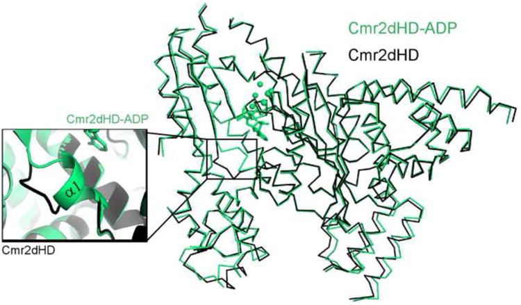
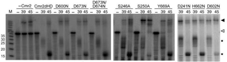
Cmr2 is the largest and an essential subunit of a CRISPR RNA-Cas protein complex (the Cmr complex) that cleaves foreign RNA to protect prokaryotes from invading genetic elements. Cmr2 is thought to be the catalytic subunit of the effector complex because of its N-terminal HD nuclease domain. Here, however, we report that the HD domain of Cmr2 is not required for cleavage by the complex in vitro. The 2.3Å crystal structure of Pyrococcus furiosus Cmr2 (lacking the HD domain) reveals two adenylyl cyclase-like and two α-helical domains. The adenylyl cyclase-like domains are arranged as in homodimeric adenylyl cyclases and bind ADP and divalent metals. However, mutagenesis studies show that the metal- and ADP-coordinating residues of Cmr2 are also not critical for cleavage by the complex. Our findings suggest that another component provides the catalytic function and that the essential role by Cmr2 does not require the identified ADP- or metal-binding or HD domains in vitro.


Figures

Figure 1
Figure 1
The RNA silencing pathway mediated by the Cmr2-containing Cmr complex in Pyrococcus furiosus. (A) The Pyrococcus furiosus Cas protein locus #1. The processing endoribonuclease Cas6 is shown in grey and the Cmr proteins are shown in blue. Cmr2 is the largest of the six Cmr proteins and is a member of the Cas10 superfamily of CRISPR polymerase proteins. (B) Schematic of crRNA processing, assembly into the Cmr RNP complex and guided cleavage of a target RNA. The pre-crRNA transcript is initially processed by Cas6 within the repeat sequence to form intermediate crRNAs which contain 8 nt of the repeat at the 5′ end (called the tag; shown in black) and the remaining 22 nt of repeat at the 3′ end. These intermediate crRNAs are then processed by an unknown mechanism into mature crRNAs, which retain the 5′ tag sequence upstream of 31 or 37 nts of the spacer sequence. The mature crRNAs are loaded on the Cmr effector complex and guide the recognition and cleavage of target RNAs through base-pairing. The Cmr complex cleaves the target at the nucleotide opposite to that 14nt from the 3′ end of the crRNA. (C) In vitro cleavage activity of the reconstituted P. furiosus Cmr complex containing the full-length Cmr2 or an N-terminal domain truncated mutant, Cmr2dHD. The Cmr complex (1 μM) containing full-length Cmr2 (wt), no Cmr2 (-Cmr2) or Cmr2 lacking the N-terminal HD domain (Cmr2dHD) was incubated with either the 39 or 45 nucleotide crRNA, followed by addition of radiolabeled target RNA (indicated by an open arrow). – lanes contain no crRNA or proteins. The 39 nt crRNA guides cleavage that results in a 20 nt cleavage product while the 45 nt crRNA results in a 14 nt cleavage product (indicated by asterisks). crRNA/target RNA duplexes are indicated by closed arrows. See also Figure S1.
Figure 2
Figure 2
Crystal structure of P. furiosus Cmr2dHD. (A) Overview and domain structure of Cmr2dHD. Cmr2dHD is a flat, triangular, four-domain protein. The N-terminal domain D1 (blue) contains a ferredoxin-like fold (which contains a P-loop) and a cysteine cluster. The D3 domain (orange) contains a ferredoxin fold that includes a conserved GGDD motif on a β-hairpin and a P-loop structure. Domains D2 (green) and D4 (red) are exclusively α-helical. (B) Topology of the Cmr2dHD structure with the same coloring scheme as panel A. Arrows represent β-sheets and cylinders represent α-helices. Dashed lines represent disordered regions. The P-loops of D1 and D3, the β-hairpin of D3 and the cysteine cluster of D1 are indicated. (C) Cmr2dHD contains two homologous ferredoxin folds (colored in blue and orange for the D1 and D3 domains respectively). The D1 ferredoxin-like fold lacks the β-hairpin but maintains the P-loop (purple). The D3 domain contains both the β-hairpin (yellow) and P-loop (purple). (D) D1 and D3 domains are homologous. The two domains have a root-means-square-deviation of 2.6 Å. (E) Electrostatic surface potential of Cmr2dHD bound with adenosine diphosphate (ADP). See also Figure S4.
Figure 3
Figure 3
Structure of Cmr2dHD bound with adenosine diphosphate (ADP) and Ca2+, and structure comparison with Mycobacterium tuberculosis adenylyl cyclase (Mt Ac). (A) Cmr2dHD binds to ADP (stick model) at its interface between D1 and D3 domain. Cyan spheres represent bound Ca2+ ions and red spheres represent water oxygen atoms. (B) Close-up view of ligand binding site of Cmr2dHD. ADP interacts with the P-loops (purple) of the D1 domain and the bound Ca2+ atoms interact with the β-hairpin (yellow) and P-loop (purple) of domain D3. (C) Crystal structure of the AB dimer (colored in blue and orange respectively) of the Mycobacterium tuberculosis (Mt) Rv1900c adenylyl cyclase bound with α, β-methyleneadenosine 5′-triphosphate (AMPCPP) ((Sinha et al., 2005), 1YBU). The Mt adenylyl cyclase dimer is displayed with its monomer A (orange) in the same orientation as D3 domain of Cmr2dHD. (D) Close-up view of ligand binding site of Mt adenyly cyclase. AMPCPP interacts primarily with the β-hairpin (yellow) and P-loop (purple) of monomer A in Mt adenylyl cyclase.
Figure 4
Figure 4
Overlay of the ADP-bound (green) and unbound (black) structures of Cmr2dHD. Conformational change is restricted to widening of the distance between the lower part of the D1 and D2 domains and the formation of the α1 helix at the nucleotide binding site (inset).
Figure 5
Figure 5
Differences in interaction of the conserved residues of Cmr2dHD and Mt adenylyl cyclase with nucleotide and metal ions. (A) Multiple sequence alignment of the most conserved portion of selected Cmr2 proteins across archaea and bacteria. Secondary structures are colored in the same scheme as in Figure 2 and are shown above the alignment (cylinders for α-helices, arrows for β-strands and lines for loops). Levels of conservation in amino acids are indicated by shades of blue with darker blue corresponding to greater conservation. The locations of the P-loop and β-hairpins are indicated. Residues that are mutated in Figure 6 or S3 are indicated by asterisks. (B) Detailed interactions at the ligand binding site of Pf Cmr2dHD. Residues within 3.5 Å of the bound ligands are displayed using the same color scheme as in Figure 2. Cyan spheres represent bound Ca2+ ions and red spheres represent water oxygen atoms. (C) Detailed interactions of Mt adenylyl cyclase active site. Residues are similarly identified and the purple sphere represents bound Mn2+. See also Figures S2, S3 and S5.
Figure 6
Figure 6
Mutational analysis of the nucleotide and metal-binding residues of Cmr2dHD. ReconstitutedCmr complexes (Cmr1-6, 1 μ M) containing no Cmr2 (-Cmr2), Cmr2dHD or mutants thereof (asindicated) were incubated with crRNAs (0.1 μ M) of 39 nt or 45 nt (as indicated) for 30 minutesbefore the addition of γ 32P-labeled target RNA (labeled by an open arrow). The reactionmixtures were incubated for 1 hour and analyzed on 15% denaturing polyacrylamide gel. – lanescontain no crRNA or proteins. Cleavage products are indicated by asterisks. Target RNA/crRNA duplexes are indicated by a closed arrow. See also Table S1.

Comment in

Similar articles

Cited by

References

    1. Adams PD, Afonine PV, Bunkoczi G, Chen VB, Davis IW, Echols N, Headd JJ, Hung LW, Kapral GJ, Grosse-Kunstleve RW, et al. PHENIX: a comprehensive Python-based system for macromolecular structure solution. Acta Crystallogr D Biol Crystallogr. 2010;66:213–221. - PMC - PubMed
    1. Anantharaman V, Iyer LM, Aravind L. Presence of a classical RRM-fold palm domain in Thg1-type 3′- 5′nucleic acid polymerases and the origin of the GGDEF and CRISPR polymerase domains. Biol Direct. 2010;5:43. - PMC - PubMed
    1. Artymiuk PJ, Poirrette AR, Rice DW, Willett P. A polymerase I palm in adenylyl cyclase? Nature. 1997;388:33–34. - PubMed
    1. Barrangou R, Fremaux C, Deveau H, Richards M, Boyaval P, Moineau S, Romero DA, Horvath P. CRISPR provides acquired resistance against viruses in prokaryotes. Science. 2007;315:1709–1712. - PubMed
    1. Beloglazova N, Petit P, Flick R, Brown G, Savchenko A, Yakunin AF. Structure and activity of the Cas3 HD nuclease MJ0384, an effector enzyme of the CRISPR interference. EMBO J. 2011;30:4616–4627. - PMC - PubMed

Publication types

MeSH terms

Substances

LinkOut - more resources