

Review
doi: 10.1016/j.cmet.2011.12.018.
FAT SIGNALS--lipases and lipolysis in lipid metabolism and signaling
Affiliations
- PMID: 22405066
- PMCID: PMC3314979
- DOI: 10.1016/j.cmet.2011.12.018
Item in Clipboard
Review
FAT SIGNALS--lipases and lipolysis in lipid metabolism and signaling
Cell Metab.
.
Abstract
Lipolysis is defined as the catabolism of triacylglycerols stored in cellular lipid droplets. Recent discoveries of essential lipolytic enzymes and characterization of numerous regulatory proteins and mechanisms have fundamentally changed our perception of lipolysis and its impact on cellular metabolism. New findings that lipolytic products and intermediates participate in cellular signaling processes and that "lipolytic signaling" is particularly important in many nonadipose tissues unveil a previously underappreciated aspect of lipolysis, which may be relevant for human disease.
Copyright © 2012 Elsevier Inc. All rights reserved.
Figures
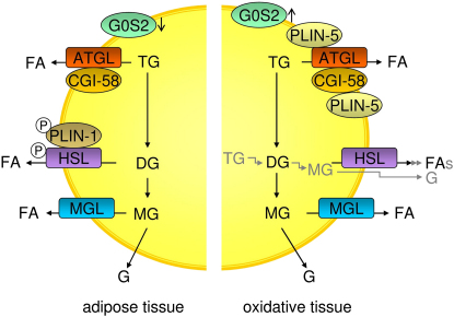
Lipolysis in Adipose and Oxidative Tissues during Fasting In adipose tissues, beta-adrenergic stimulation of lipolysis leads to the consecutive hydrolysis of TG and the formation of FAs and glycerol. The process requires three enzymes: ATGL cleaves the first esterbond in TGs, HSL hydrolyzes DGs, and MGL MGs. For full hydrolytic activity, ATGL interacts with its coactivator protein CGI-58, whereas HSL is phosphorylated, translocates to the LD, and interacts with phosphorylated PLIN-1. Expression of the ATGL inhibitor G0S2 during fasting is low in adipose and high in oxidative tissues (e.g., liver). In oxidative tissues PLIN-1 is not present on LDs. Instead, PLIN-5 is expressed and interacts with both ATGL and CGI-58, facilitating LD localization of these proteins. ATGL, adipose triglyceride lipase; CGI-58, comparative gene identification-58; DG, diacylglycerol; FA, fatty acid; G, glycerol; G0S2, G0/G1 switch gene 2; HSL, hormone-sensitive lipase; MG, monoacylglycerol; MGL, monoglyceride lipase; PLIN-1, perilipin-1; PLIN-5, perilipin-5; TG, triacylglycerol.
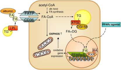
ATGL-Mediated Lipolysis Is Required for PPAR Signaling and OXPHOS Fatty acids from exogenous or endogenous sources are activated to acyl-CoAs, which are subject to mitochondrial oxidation or TG formation. ATGL-mediated lipolysis of TG generates lipolytic products (FA and DG), which may act directly (e.g., FA) or after conversion (e.g., DG to phospholipids) as ligands for nuclear receptors (for details see text). Activation of nuclear receptor PPARα via lipolytic cleavage of TGs is required for normal mitochondrial function and OXPHOS. In ATGL-deficient mice, defective PPARα activation and OXPHOS can be restored by treatment with PPARα agonists. ATGL, adipose triglyceride lipase; CD36, cluster of differentiation 36; DG, diacylglycerol; FA, fatty acid; FATP, fatty acid transport protein; LPL, lipoprotein lipase; OXPHOS, oxidative phosphorylation; PPARα/δ, peroxisome proliferator-activated receptor alpha/delta; RA, retinoic acid; RXR, retinoid X receptor; TG, triacylglycerol.
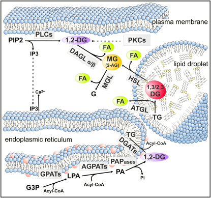
Lipolysis and Lipid Signaling Lipid intermediates involved in cellular signaling are generated by anabolic and catabolic reactions in distinct cellular compartments. 1,2-DGs, the ligands of conventional and novel PKCs, are formed at the plasma membrane by PLC-mediated degradation of PIP2. This reaction also generates IP3, a signaling molecule, which leads to Ca2+ efflux from the ER. De novo synthesis of 1,2-DGs at the ER may also contribute to PKC activation. FAs are ligands for nuclear receptors. They are generated by de novo synthesis or hydrolysis of neutral lipids or phospholipids. 2-AG is an important MG involved in endocannabinoid signaling. It originates from membrane-associated phospholipid hydolysis by PLCs and the subsequent hydrolysis of DGs by DAGLs. The contribution of TG hydrolysis by ATGL and HSL to cellular 2-AG concentrations is not known. The 2-AG signal is inactivated by MGL. AGPAT, acyl-CoA acylglycerol-3-phosphate acyltransferase; 2-AG, 2-arachidonoyl-glycerol; ATGL, adipose triglyceride lipase; DAGL, diacylglycerol lipase; DG, diacylglycerol; 11, 2-DG, diacyl-sn1,2-glycerol, DGAT, acyl-CoA: diacylglycerol acyltransferase; FA, fatty acid; G, glycerol; G3P, glycerol-3-phosphate; GPAT, glycerol-3-phosphate acyltransferase; HSL, hormone-sensitive lipase; IP3, inositol-1,4,5-trisphosphate; LPA, lysophosphatidic acid; MG, monoacylglycerol; MGL, monoglyceride lipase; PA, phosphatidic acid; PAPase, PA phosphohydrolase; PIP2, phosphatidylinositol 4,5-bisphosphate; PKC, protein kinase C; PLC, phospholipase C; TG, triacylglycerol.
References
-
- Agustsson T., Rydén M., Hoffstedt J., van Harmelen V., Dicker A., Laurencikiene J., Isaksson B., Permert J., Arner P. Mechanism of increased lipolysis in cancer cachexia. Cancer Res. 2007;67:5531–5537. - PubMed
-
- Anthonsen M.W., Rönnstrand L., Wernstedt C., Degerman E., Holm C. Identification of novel phosphorylation sites in hormone-sensitive lipase that are phosphorylated in response to isoproterenol and govern activation properties in vitro. J. Biol. Chem. 1998;273:215–221. - PubMed
Publication types
MeSH terms
Substances
Grants and funding
LinkOut - more resources
Full Text Sources
Other Literature Sources
