. 2012 Mar 9;90(3):558-64.
doi: 10.1016/j.ajhg.2012.02.006.

Exome sequencing reveals mutations in TRPV3 as a cause of Olmsted syndrome

Affiliations

Exome sequencing reveals mutations in TRPV3 as a cause of Olmsted syndrome

Zhimiao Lin et al. Am J Hum Genet. .

Abstract

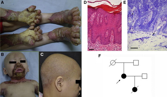
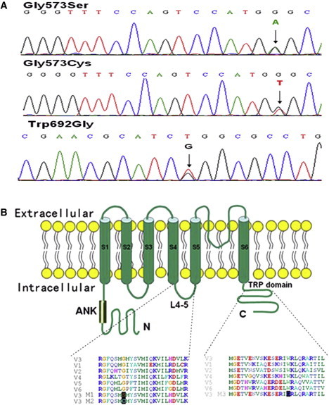
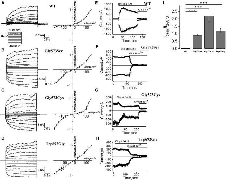
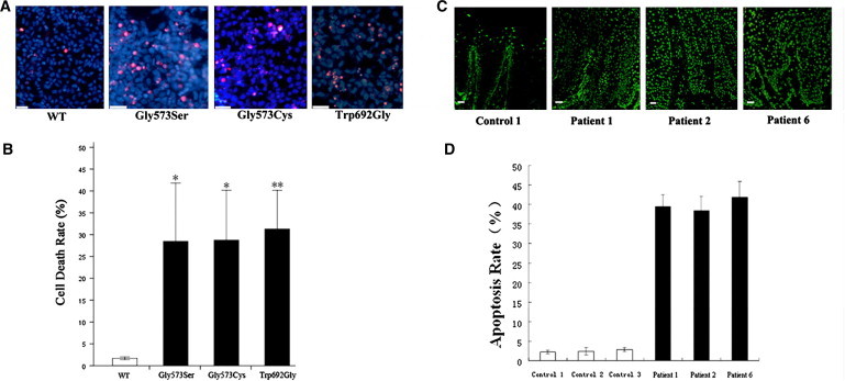
Olmsted syndrome (OS) is a rare congenital disorder characterized by palmoplantar and periorificial keratoderma, alopecia in most cases, and severe itching. The genetic basis for OS remained unidentified. Using whole-exome sequencing of case-parents trios, we have identified a de novo missense mutation in TRPV3 that produces p.Gly573Ser in an individual with OS. Nucleotide sequencing of five additional affected individuals also revealed missense mutations in TRPV3 (which produced p.Gly573Ser in three cases and p.Gly573Cys and p.Trp692Gly in one case each). Encoding a transient receptor potential vanilloid-3 cation channel, TRPV3 is primarily expressed in the skin, hair follicles, brain, and spinal cord. In transfected HEK293 cells expressing TRPV3 mutants, much larger inward currents were recorded, probably because of the constitutive opening of the mutants. These gain-of-function mutations might lead to elevated apoptosis of keratinocytes and consequent skin hyperkeratosis in the affected individuals. Our findings suggest that TRPV3 plays essential roles in skin keratinization, hair growth, and possibly itching sensation in humans and selectively targeting TRPV3 could provide therapeutic potential for keratinization or itching-related skin disorders.


Figures

Figure 1
Figure 1
Phenotypic Characteristics of OS (A) Bilateral mutilating palmoplantar keratoderma, flexion deformities and constriction of digits in individual 1. (B) Keratotic plaques involving periorificial, neck and axillary areas in individual 1. (C) Diffused alopecia with follicular papules in individual 2. (D) Skin biopsy section demonstrating psoriasiform hyperplasia, orthohyperkeratosis and parakeratosis in individual 1 (hematoxylin-eosin staining; the scale bar represents 100 μm). (E) Individual 1, skin biopsy section showing profound mast cell infiltration in upper dermis (toluidine blue staining; the scale bar represents 100 μm). (F) The pedigree of individual 3 (arrow indicates individual 3).
Figure 2
Figure 2
Mutations and Schematic Structure of TRPV3 (A)Sequencing results demonstrating heterozygous TRPV3 mutation of c.1717G>A (amino acid change of p.Gly573Ser) in individuals 1, 3, 4, and 6; c.1717G>T (p.Gly573Cys) in individual 2; and c.2074T>G (p.Trp692Gly) in individual 5. (B) Schematic structure of TRPV3 protein and multiple alignment of the linker region between S4 and S5 and the TRP domain, with the alignments of corresponding segments of human TRPV1-2 and 4-6 proteins. Gly573 and Trp692 are highly conserved residues. The mutation sites that produce p.Gly573Ser (M1), p.Gly573Cys (M2), and p.Trp692Gly (M3) are also aligned with the substitution positions (black highlighted).
Figure 3
Figure 3
Whole-Cell and Inside-Out Recordings of TRPV3 Currents from Transfected HEK293 Cells (A–D) Representative whole-cell current traces in HEK293 cells expressing TRPV3 or mutant channels in response to the voltage step protocol (left panels), and respective current-voltage plots from the steady-state currents (right panels). (E–H) Representative currents from inside-out patches in HEK293 cells expressing TRPV3 or mutant channels in response to stimulation by 300 μM 2-APB or inhibition by 130 mM Ba2+ to assess the level of leak currents, at −80 mV and +80 mV. WT TRPV3 current is activated by 2-APB and followed by a characteristic decay. Note that the mutants Gly573Ser, Gly573Cys, and Trp692Gly show little activation, then inhibition in the presence of 2-APB, and a robust block by Ba2+. (I) Comparison of ratios of currents evoked by 2-APB (300 μM) over basal currents between the TRPV3 WT and mutant channels. (t test, n = 3–7. p = 0.00001). Error bars represent SEM.
Figure 4
Figure 4
Cell Death of Transfected HEK293 Cells and Apoptosis of Affected Individuals' Keratinocytes (A) Merged images of transfected HEK293 cells stained with PI (red) and Hoechst 33342 (blue). Mutants show significantly more PI-positive cells compared to wild-type TRPV3. (B) Quantification of cell death rates in transfected HEK293 cells (red nuclei/blue nuclei × 100%). Data are averaged from three independent experiments. p < 0.05, ∗∗p < 0.01. (C) Fluorescence microscope images (TUNEL method) of palm skin biopsy sections from an unrelated control, individuals 1, 2, and 6. Large amount of apoptotic cells (with fluorescent nuclei) are seen in individuals with OS. The scale bars represent 50 μm. (D) Quantification of apoptotic keratinocytes in the skin sections. The mean proportion of apoptotic cells are significantly higher in the group of OS patients than in the group of normal controls (t test, n = 3, p = 0.000005). Error bars represent SEM.

References

    1. Olmsted H.C. Keratodermia palmaris et plantaris congenitalis: Report of a case showing associated lesions of unusual location. Am. J. Dis. Child. 1927;33:757–764.
    1. Mevorah B., Goldberg I., Sprecher E., Bergman R., Metzker A., Luria R., Gat A., Brenner S. Olmsted syndrome: mutilating palmoplantar keratoderma with periorificial keratotic plaques. J. Am. Acad. Dermatol. 2005;53(5, Suppl 1):S266–S272. - PubMed
    1. Ogawa F., Udono M., Murota H., Shimizu K., Takahashi H., Ishida-Yamamoto A., Iizuka H., Katayama I. Olmsted syndrome with squamous cell carcinoma of extremities and adenocarcinoma of the lung: failure to detect loricrin gene mutation. Eur. J. Dermatol. 2003;13:524–528. - PubMed
    1. Vosynioti V., Kosmadaki M., Tagka A., Katsarou A. A case of Olmsted syndrome. Eur. J. Dermatol. 2010;20:837–838. - PubMed
    1. Tharini G.K., Hema N., Jayakumar S., Parveen B. Olmsted syndrome: report of two cases. Indian J. Dermatol. 2011;56:591–593. - PMC - PubMed

Publication types