. 2012;21(6):1121-35.
doi: 10.3727/096368911X627381. Epub 2012 Mar 8.

Magnetic enhancement of cell retention, engraftment, and functional benefit after intracoronary delivery of cardiac-derived stem cells in a rat model of ischemia/reperfusion

Affiliations

Magnetic enhancement of cell retention, engraftment, and functional benefit after intracoronary delivery of cardiac-derived stem cells in a rat model of ischemia/reperfusion

Ke Cheng et al. Cell Transplant. 2012.

Abstract

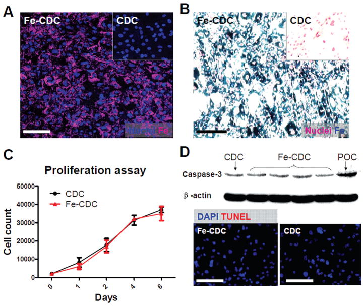
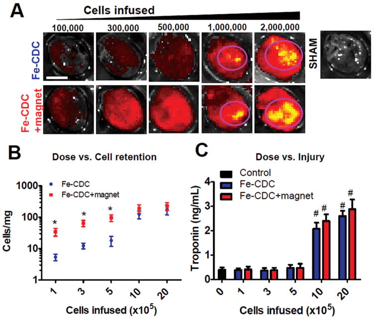
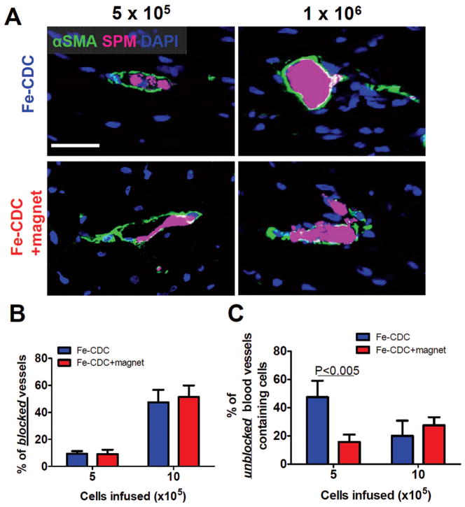
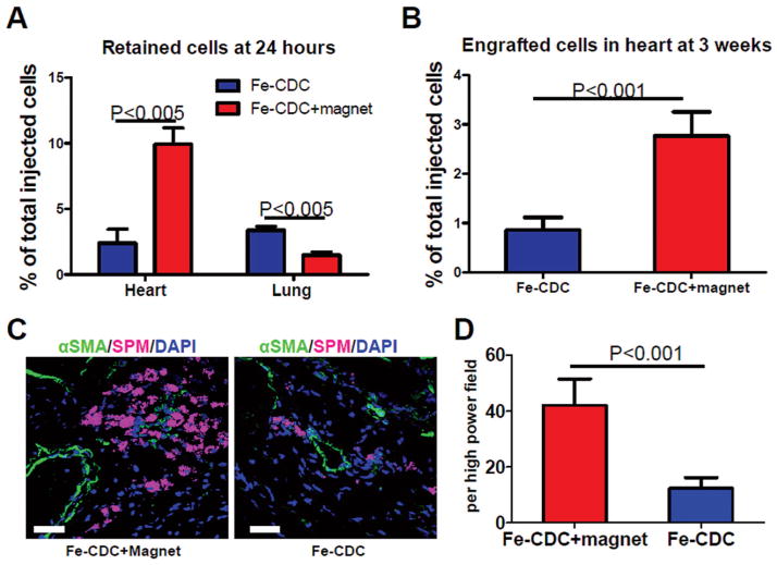
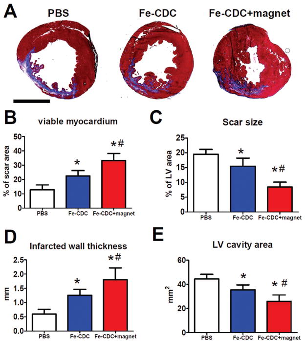
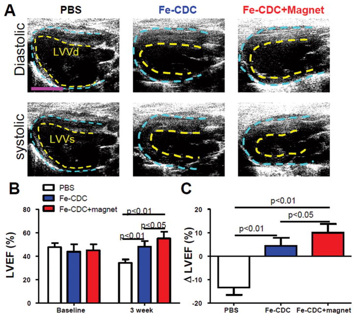
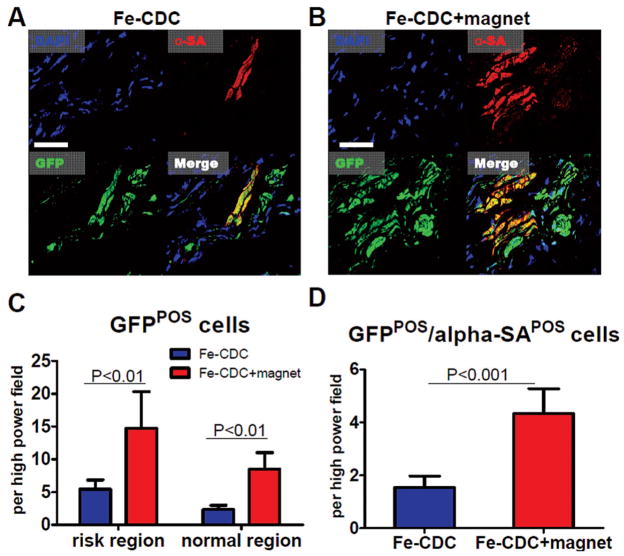
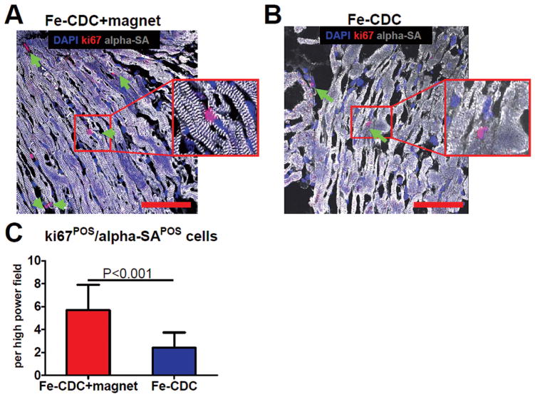
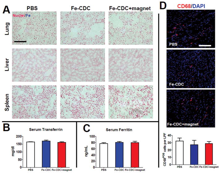
The efficiency of stem cell transplantation is limited by low cell retention. Intracoronary (IC) delivery is convenient and widely used but exhibits particularly low cell retention rates. We sought to improve IC cell retention by magnetic targeting. Rat cardiosphere-derived cells labeled with iron microspheres were injected into the left ventricular cavity of syngeneic rats during brief aortic clamping. Placement of a 1.3 Tesla magnet ~1 cm above the heart during and after cell injection enhanced cell retention at 24 h by 5.2-6.4-fold when 1, 3, or 5 × 10(5) cells were infused, without elevation of serum troponin I (sTnI) levels. Higher cell doses (1 or 2 × 10(6) cells) did raise sTnI levels, due to microvascular obstruction; in this range, magnetic enhancement did not improve cell retention. To assess efficacy, 5 × 10(5) iron-labeled, GFP-expressing cells were infused into rat hearts after 45 min ischemia/20 min reperfusion of the left anterior coronary artery, with and without a superimposed magnet. By quantitative PCR and optical imaging, magnetic targeting increased cardiac retention of transplanted cells at 24 h, and decreased migration into the lungs. The enhanced cell engraftment persisted for at least 3 weeks, at which time left ventricular remodeling was attenuated, and therapeutic benefit (ejection fraction) was higher, in the magnetic targeting group. Histology revealed more GFP(+) cardiomyocytes, Ki67(+) cardiomyocytes and GFP(-)/ckit(+) cells, and fewer TUNEL(+) cells, in hearts from the magnetic targeting group. In a rat model of ischemia/reperfusion injury, magnetically enhanced intracoronary cell delivery is safe and improves cell therapy outcomes.


Conflict of interest statement

The authors declare no conflict of interest.

Figures

Figure 1
Figure 1
Superparamagnetic microsphere (SPM) labeling of rat cardiosphere-derived cells (CDCs). (A) Rat CDCs were coincubated with flash red-conjugated SPMs for 24 h at a 500:1 SPM/cell ratio. CDCs were then examined by fluorescence microscopy. (B) Cells were fixed, stained for Prussian blue (iron), and counterstained with nuclear red. Nonlabeled cells did not express flash red fluorescence or Prussian blue staining (A & B, insets). (C) Cell Counting Kit-8 (CCK-8) proliferation assay of CDCs and Fe-CDCs (n = 3). No significant differences were detected. (D) Western blot analysis of caspase-3 and terminal deoxynucleotidyl transferase dUTP nick end labeling (TUNEL) staining revealed no evident increase of apoptosis in the Fe-CDCs. DAPI = 4′,6-diamidino-2-phenylindole. Scale Bars: 50 μm.
Figure 2
Figure 2
Dosage optimization for IC infusion of Fe-CDCs with and without magnetic enhancement. Animals were sacrificed 24 h after cell infusion. Hearts were excised for fluorescent imaging and qPCR quantification of cell numbers. Serum samples were obtained for measurement of troponin I (TnI) as an indicator of myocardial injury. (A) Fluorescent imaging revealed escalation of fluorescent intensity with increasing cell infusion doses in both groups. High density areas were seen when 1 × 10⁶ and 2 × 10⁶ cells were infused (circled with pink). Control hearts (infused with PBS) exhibited no fluorescence. The color ranges were set the same for all images. (B) Cell numbers per milligram of heart tissue were measured by qPCR and plotted against doses (n = 3 per data point). *p < 0.05 when compared to the Fe-CDC group. (C) Serum TnI values were measured by ELISA and plotted against doses (n = 3 per data point). #p < 0.05 when compared to the control (dose = 0) group. Scale bar: 5 mm.
Figure 3
Figure 3
Microembolization of CDCs at high cell infusion doses. Animals were sacrificed 24 h after IC infusion and representative heart sections were stained for α-smooth muscle actin (α-SMA) to detect blood vessels. Fe-CDCs were visualized by flash red fluorescence. (A) At the infusion dose of 5 × 10⁵ Fe-CDCs, blood vessels containing cells were readily detected and the vessels were still patent; at the dose of 1 × 10⁶ Fe-CDCs, many blood vessels were blocked by cell clumps. (B) Quantification of blocked vessels. (C) Quantification of unblocked vessels that contain cells. n = 3 animals per group. Scale bar: 50 μm.
Figure 4
Figure 4
Effects of magnetic targeting on short-term cell retention and long-term cell engraftment. (A) Female animals (n = 5) were sacrificed 24 h after cell injection. Donor male cells persistent in the female hearts were detected by quantitative PCR for the sex-determining region Y (SRY) gene. (B) Similar PCR experiment performed 3 weeks after injection. Ischemia/reperfusion animals that received 500,000 Fe-CDCs with and without magnetic enhancement were sacrificed 72 h after cell infusion (n = 3 per group). Representative heart sections were stained for α-SMA for blood vessels. Fe-CDCs were visualized with flash red fluorescence. (C) Representative confocal images from the Fe-CDC + magnet and Fe-CDC group. (D) Quantification of Fe-CDCs per high power field. Scale bar: 50 μm.
Figure 5
Figure 5
Morphometric heart analysis. (A) Representative Masson’s trichrome-stained myocardial sections 3 weeks after treatment (n = 7 per group). Scar tissue and viable myocardium are identified by blue and red color, respectively. (B–E) Quantitative analysis and left ventricle (LV) morphometric parameters. *p < 0.05 when compared to control. #p < 0.05 when compared to the Fe-CDC group. Scale bar: 5 mm.
Figure 6
Figure 6
Magnetic targeting enhances functional benefit of IC delivery of Fe-CDCs. (A) Representative long-axis diastolic and systolic images at 3 weeks after treatment. The pericardium and endocardium are outlined with yellow and blue dotted lines, respectively. LVVd, left ventricular volume in diastole; LVVs, left ventricular volume in systole. (B) Left ventricular ejection fraction (LVEF) measured by echocardiography at baseline and 3 weeks after cell injection (n = 9 per group). Baseline LVEFs were indistinguishable among the three groups. (C) Changes of LVEF from baseline to 3 weeks in each group. Values are expressed as mean ± SD. Scale var: 5 mm.
Figure 7
Figure 7
Engraftment and cardiac differentiation of infused CDCs. Animals were sacrificed 3 weeks after cell infusion (n = 5 animals per group) and representative heart sections were stained for DAPI, green fluorescent protein (GFP), α-sarcomeric actin (α-SA). (A, B) Representative confocal images from the Fe-CDC and Fe-CDC + magnet group. The Fe-CDC + magnet group had more GFP+ and GFP+ /αSA+ cells. This indicated that magnetic targeting improved long-term cell engraftment. (C) Quantification of GFP+ cells in the risk and normal region. (D) Quantification of GFP+ /α-SA+ cells. Scale bar: 100 μm.
Figure 8
Figure 8
Ki67-positive cardiomyocytes. Animals were sacrificed 3 weeks after cell infusion (n = 5 animals per group) and representative heart sections were stained for DAPI, Ki67, and α-sarcomeric actin (α-SA). (A, B) Representative confocal images from the Fe-CDC and Fe-CDC + magnet group. The Fe-CDC + magnet group had more Ki67+ /α-SA+ cells (green arrows and insets), indicating more cardiomyocytes were proliferative or newly formed. (C) Quantification of Ki67+ /α-SA+ cells. Scale bars: 100 μm.
Figure 9
Figure 9
Toxicity of iron administration. (A) Prussian blue staining revealed no evident iron clusters from the lungs, livers, and spleens in all three groups. No iron overdose caused by iron labeling and/or magnetic enhancement. Serum samples were obtained 3 weeks after treatment. Transferrin (B) and ferritin (C) levels were measured by ELISA. The values from all three treatment groups were indistinguishable (n = 3 animals per group). Tissue densities of CD68+ macrophages were identical among all treatment groups. Representative confocal images showing the presence of CD68+ macrophages from hearts excised at 3 weeks from the PBS control, Fe-CDC, and Fe-CDC + magnet groups. (D) Quantification of total CD68+ macrophages per low power field (LPF; n = 5 animals per group). The values from all three treatment groups were indistinguishable. Scale bars: 100 μm.

References

    1. Al Kindi A, Ge Y, Shum-Tim D, Chiu RC. Cellular cardiomyoplasty: Routes of cell delivery and retention. Front Biosci. 2008;13:2421–2434. - PubMed
    1. Arbab AS, Bashaw LA, Miller BR, Jordan EK, Lewis BK, Kalish H, Frank JA. Characterization of biophysical and metabolic properties of cells labeled with superparamagnetic iron oxide nanoparticles and transfection agent for cellular mr imaging. Radiology. 2003;229:838–846. - PubMed
    1. Arbab AS, Jordan EK, Wilson LB, Yocum GT, Lewis BK, Frank JA. In vivo trafficking and targeted delivery of magnetically labeled stem cells. Hum Gene Ther. 2004;15:351–360. - PubMed
    1. Assis AC, Carvalho JL, Jacoby BA, Ferreira RL, Castanheira P, Diniz SO, Cardoso VN, Goes AM, Ferreira AJ. Time-dependent migration of systemically delivered bone marrow mesenchymal stem cells to the infarcted heart. Cell Transplant. 2010;19:219–230. - PubMed
    1. Bartunek J, Sherman W, Vanderheyden M, Fernandez-Aviles F, Wijns W, Terzic A. Delivery of biologics in cardiovascular regenerative medicine. Clin Pharmacol Ther. 2009;85:548–552. - PubMed

Publication types

MeSH terms