

doi: 10.1186/1741-7007-10-19.
What does the concept of the stem cell niche really mean today?
Affiliations
- PMID: 22405133
- PMCID: PMC3298504
- DOI: 10.1186/1741-7007-10-19
Item in Clipboard
What does the concept of the stem cell niche really mean today?
BMC Biol.
.
No abstract available
Figures
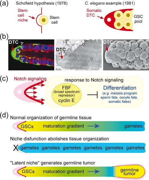
The Caenorhabditis elegans distal tip cell (DTC) and the concept of a stem cell niche. (a) Left, the stem cell niche hypothesis from Schofield [1]; right, the C. elegans DTC (red) provides the stem cell niche for the germline stem cell (GSC) pool (yellow). (b) Images of the adult DTC and its processes. Left, cytoplasmic green fluorescent protein (green) highlights the DTC and its processes that embrace GSCs. Blue, germline nuclei; red, germline membranes. Modified from [10]. Middle, electron microscopy (EM) image of DTC and its processes. Modified from [10]. Right, scanning EM image of distal gonad; image courtesy of David Greenstein [19]. An asterisk (*) marks one GSC in each image; arrowheads mark processes. (c) Molecular view of the niche and its control of GSC self-renewal or differentiation. Dark red, minimalist view of niche as the surface presenting Notch ligands; pink, broader view of niche including DTC as integral to providing the microenvironment for GSC control. (d) Expansion of niche concept based on investigations of DTC and Notch signaling in C. elegans. See text for explanation.
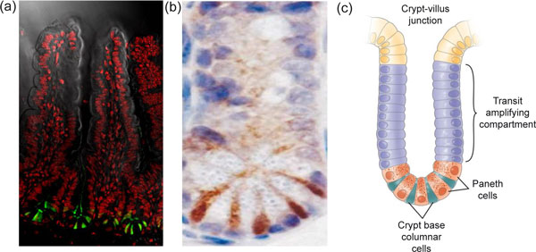
Expression of Lgr5-GFP at crypt bottoms. (a) Lgrf-5 is shown in green, with a counterstain for DNA in red to outline crypts and villi. (b) Lgr5 marks cycling crypt base columnar cells. Lgr5 expression appears in brown, in between the white/blue Paneth cells at crypt bottoms. (c) Schematic of crypt architecture. Reproduced, with permission from Elsevier, from Barker N, Clevers H: Gastroenterology 2007, 133:1755-1760.
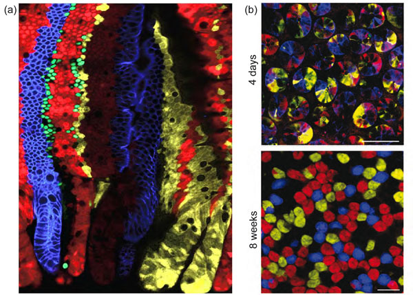
Stem cells are marked in individual colors by the multicolor Cre reporter Confetti. (a) Each crypt becomes monochromatic over time (bottom of image), producing parallel bands of differently colored cells on villus flanks. (b) Confocal sectioning through multiple crypt bottoms. When individual stem cells are marked with different Confetti colors, crypts resolve to monoclonality (that is, they become monochromatic in 4 to 8 weeks due to neutral competition of the stem cells). All images in this figure were reproduced with permission from Elsevier, from Snippert H et al.: Cell 2010, 143:134-144.

Homeostasis and repair of the adult tissues depends on tissue-specific stem cells. (a) The architecture of the hair follicle stem cell niche. The hair follicle stem cells are marked by CD34 staining (in green). One of their important niche components is the inner layer of the bulge, marked by K6 staining (in red) and composed of differentiated hair follicle stem cell progeny that underwent the transition from slow-cycling to faster-cycling. This feature was exploited by bromodeoxyuridine (BrdU) nucleotide pulse-chase to mark the inner layer cells with blue BrdU staining in the figure. This inner layer of bulge cells plays a key role in maintaining the quiescence of the outer layer of hair follicle stem cells. This image is courtesy of Y-C Hsu and E Fuchs. (b) The hair follicle stem cells are marked by CD34 staining (in green) and are quiescent, due to the high level of bone morphogenetic protein (BMP) signaling within the niche, as shown here by the nuclear staining for phosphorylated Smad1 (in red), the transcriptional effector of the BMP pathway. The nuclei of the skin cells are marked here in Keratin-5 (blue), which reveals the presence of the emerging hair follicle below the activated stem cell niche. This is a classical sign of entry into the growth phase of the new hair cycle. This image is courtesy of N Oshimori and E Fuchs.
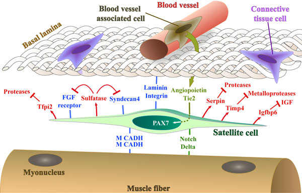
A representation (not to scale) of the satellite cell, marked by Pax7, in its niche on the muscle fiber under the basal lamina in proximity to a blood vessel. Cell adhesion molecules, signals received from surrounding tissues, and molecules secreted by the satellite cell that regulate the niche and promote the quiescent state, discussed in the text, are illustrated. IGF, insulin-like growth factor; M CADH, M-cadherin.
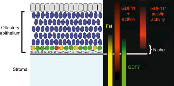
Model in which molecular gradients along the apical-basal axis of the olfactory epithelium (OE) generate the neuronal stem cell niche. In the OE, differentiation proceeds in a basal-apical direction, with stem cells (yellow) and intermediate progenitors (shown in red and green) lying in a basal compartment, underneath the post-mitotic olfactory receptor neurons (shown in blue) to which they give rise. Note that localized expression, along with the interaction of growth differentiation factor (GDF)11 and activin with Fst (a high affinity antagonist of both proteins), create a niche within the OE in which the activity of factors that promote neurogenesis (for example, GDF7) is high, and that of factors that inhibit neurogenesis (for example, GDF11, activin) is low.
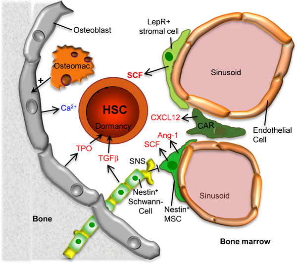
Model showing the various cell types comprising the bone marrow hematopoietic stem cell (HSC) niche. The dormant status of HSCs is maintained by transforming growth factor beta (TGF-ß) and thrombopoietin (TPO) produced by nestin+ non-myelinating Schwann cells and osteoblasts, respectively. Stem cell factor (SCF), which is essential for HSC maintenance, is mainly produced by leptin-receptor (LepR)-expressing mesenchymal stromal cells but also by nestin+ mesenchymal stem cells (MSCs) as well as sinusoidal endothelial cells (not shown). The sympathetic nervous system (SNS) negatively affects the activity of nestin+ MSCs. CXCL12 abundant reticular (CAR) cells produce the chemokine CXCL12, which facilitates lodging and engraftment as has been suggested for the high calcium concentration near the endosteal osteoblasts. The four stromal cell populations indicated in green may be somewhat overlapping and the relationship between these cell types remains to be elucidated. Osteomacs are specific macrophages that promote survival to osteoblasts and support nestin+ MSCs (not shown). Ang-1, angiopoietin-1.
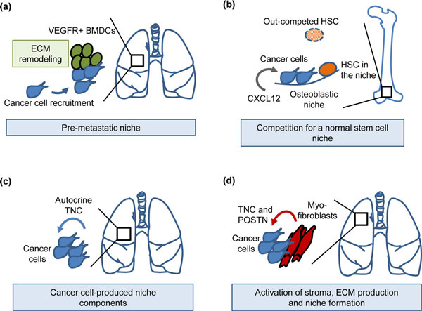
Examples of metastatic niches during early colonization of distant organs. (a) Systemic changes induced by the primary breast tumor: mobilization of VEGFR1+ bone marrow-derived cells (BMDCs), recruitment to the lungs, extracellular matrix (ECM) remodeling and formation of a pre-metastatic niche [107,108]. The pre-metastatic niche promotes the colonization of breast cancer cells in the lungs. (b) Prostate cancer cells enter the osteoblastic niche, competing with hematopoietic stem cells (HSCs) for niche interactions in bone [109]. CXCL12 chemokine promotes colonization of prostate cancer cells in the bone niche via CXCR4 interaction [111]. (c) Breast cancer cells bringing their own niche material, tenascin C (TNC), to a distant site thereby promoting early colonization of the lungs [119]. (d) Activated myofibroblasts produce the metastatic niche components TNC and periostin (POSTN), resulting in enhanced metastatic outgrowth [119-121]. VEGFR, vascular endothelial growth factor receptor.
References
-
- Schofield R. The relationship between the spleen colony-forming cell and the haemopoietic stem cell. Blood Cells. 1978;4:7–25. - PubMed
Publication types
MeSH terms
Grants and funding
LinkOut - more resources
Full Text Sources
Other Literature Sources
