. 2012 Mar 8;73(5):903-10.
doi: 10.1016/j.neuron.2012.01.018.

FLRT proteins are endogenous latrophilin ligands and regulate excitatory synapse development

Affiliations

FLRT proteins are endogenous latrophilin ligands and regulate excitatory synapse development

Matthew L O'Sullivan et al. Neuron. .

Abstract

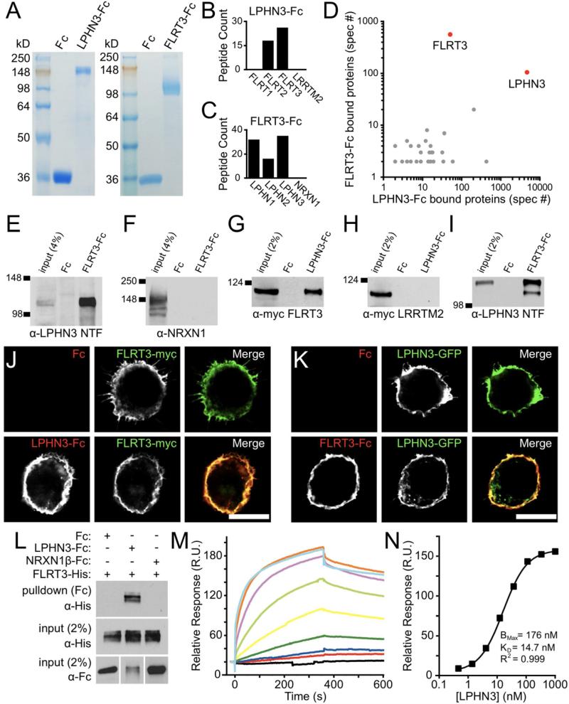
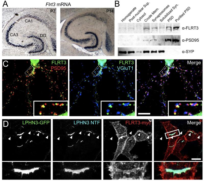
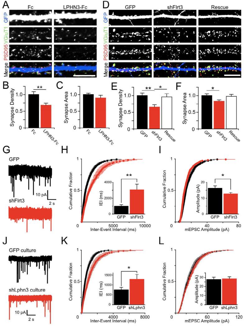
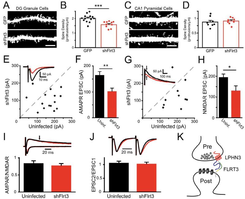
Latrophilins (LPHNs) are a small family of G protein-coupled receptors known to mediate the massive synaptic exocytosis caused by the black widow spider venom α-latrotoxin, but their endogenous ligands and function remain unclear. Mutations in LPHN3 are strongly associated with attention deficit hyperactivity disorder, suggesting a role for latrophilins in human cognitive function. Using affinity chromatography and mass spectrometry, we identify the FLRT family of leucine-rich repeat transmembrane proteins as endogenous postsynaptic ligands for latrophilins. We demonstrate that the FLRT3 and LPHN3 ectodomains interact with high affinity in trans and that interference with this interaction using soluble recombinant LPHN3, LPHN3 shRNA, or FLRT3 shRNA reduces excitatory synapse density in cultured neurons. In addition, reducing FLRT3 levels with shRNA in vivo decreases afferent input strength and dendritic spine number in dentate granule cells. These observations indicate that LPHN3 and its ligand FLRT3 play an important role in glutamatergic synapse development.


Figures

Figure 1
Figure 1. Identification of FLRT3 as a novel endogenous ligand for LPHN3
(A) Coomassie stained SDS-PAGE gels showing ecto-LPHN3-Fc (left) and ecto-FLRT3-Fc (right) purified proteins running as single bands. (B, C) Number of distinct tryptic peptides for selected proteins detected by mass spectrometry from ecto-LPHN3-Fc (B) or ecto-FLRT3-Fc (C) affinity purifications. (D) Frequency of detection of all peptides (total spectra count, spec #) for proteins identified in both LPHN3 and FLRT3 affinity purifications. (E-F) Pull-downs from whole brain extracts with ecto-FLRT3-Fc protein or control Fc protein. Ecto-FLRT3-Fc precipitates LPHN3 NTF (E), but not NRXN1 from brain (F). (G-I) Pull-downs on lysate of HEK293 cells transfected with FLRT3-myc (G), myc-LRRTM2 (H), or LPHN3-GFP (I) using ecto-Fc or control Fc proteins. Ecto-LPHN3-Fc precipitates FLRT3-myc (G), but not myc-LRRTM2 from HEK cell lysate (H). (I) Ecto-FLRT3-Fc precipitates LPHN3-GFP NTF. (J, K) Binding of soluble ecto-LPHN3-Fc (J) and ecto-FLRT3-Fc (K) proteins to HEK cells expressing FLRT3 or LPHN3. Scale bar 10 μm. (L) Direct interaction of FLRT3 and LPHN3 ectodomains. Fc, ecto-LPHN3-Fc, or ecto-NRXN1β(-S4)-Fc was mixed with FLRT3-His, precipitated, and analyzed by western blot. FLRT3-His only binds to ecto-LPHN3-Fc (top). (M) Surface Plasmon Resonance (SPR)-based measurement of ecto-LPHN3-Fc binding to immobilized ecto-FLRT3. Each colored trace represents the binding response for a different concentration of ecto-LPHN3-Fc, from 1 μM to 0.45 μM in three-fold dilutions, separated by wash and regeneration steps. (N) Concentration-response function of LPHN3-FLRT3 SPR binding. The KD of the interaction is 14.7 nM.
Figure 2
Figure 2. FLRT3 is a postsynaptic protein capable of interaction with LPHN3 in trans
(A) In situ hybridization with anti-sense Flrt3 probe in horizontal sections from P7 (left) and P14 (right) mouse brain. (B) Western blot for FLRT3 and known synaptic proteins in fractionated rat brain protein samples. Fractions, from left to right: homogenate, post-nuclear supernatant, cytosol, crude membrane, synaptosome, detergent solubilized synaptosome, postsynaptic density, and purified postsynaptic density. (C) In dissociated hippocampal neurons, FLRT3-myc (green) localizes to dendrites in a punctate fashion, and partially co-localizes with the postsynaptic marker PSD95 (red) and is closely associated with presynaptic VGluT1 puncta (blue). (D) Hippocampal neurons transfected with LPHN3-GFP were co-cultured with HEK cells expressing FLRT3-myc and transfected axons (arrowheads) were imaged where they contacted transfected HEK cells. LPHN3-GFP, LPHN3 NTF, and FLRT3-myc were all enriched at axon-HEK cell contacts. Scale bar in (C, D), 10 μm.
Figure 3
Figure 3. FLRT3 and LPHN3 regulate glutamatergic synapse number
(A) Competitive disruption of endogenous LPHN interactions with ecto-LPHN3-Fc reduces excitatory synapse density. Hippocampal cultures were fixed and immunostained for VGluT1 (green), PSD95 (red), and GFP (blue) at 14DIV after 6 days of treatment with 10 μg/ml Fc (left) or ecto-LPHN3-Fc (right). (B) LPHN3-Fc treatment reduces the density of co-localizing VGluT1 and PSD95 puncta on dendrites of identified GCs (Fc 1.00 ± 0.05, n=17; LPHN3-Fc 0.68 ± 0.06, n=14; p=0.01) (C) Synaptic puncta did not significantly differ in size between Fc- and ecto-LPHN3-Fc-treated neurons (Fc 1.00 ± 0.04, n=17; LPHN3-Fc 0.90 ± 0.08, n=14; n.s.). (D) Hippocampal neurons were electroporated with GFP control plasmid (GFP), a plasmid encoding shFlrt3 and GFP (shFlrt3), or the shFlrt3 plasmid plus an shFlrt3-resistant FLRT3-myc expression construct (Rescue), and processed for immunofluorescence as in (A). (E) Synapse density was reduced by shFlrt3 and returned to control levels in the Rescue condition (GFP 1.00 ± 0.08, n=35; shFlrt3 0.66 ± 0.06, n=33; Rescue 0.96 ± 0.09, n=34; ANOVA p<0.01, significant post-hoc comparisons shown on graph). (F) Synapse area was slightly reduced by shFlrt3 (GFP 1.00 ± 0.05, n=35; shFlrt3 0.83 ± 0.04, n=33; Rescue 0.98 ± 0.06, n=34; ANOVA p<0.05, significant post-hoc comparisons shown on graph). (G) Example mEPSC traces recorded from cultured hippocampal neurons electroporated with GFP (black) or shFlrt3 (red) plasmids. (H) Summary of mEPSC frequencies plotted as cumulative probability distributions of inter-event intervals (IEIs) for GFP (black) or shFlrt3 (red) electroporated cells. Inset: Quantification of mean mEPSC IEIs. shFlrt3 cells have longer mean IEIs than control neurons (GFP 1028 ± 147 ms, n=15; shFlrt3 3069 ± 688 ms, n=15; p < 0.01). (I) Summary of mEPSC amplitude plotted as cumulative probability distributions for GFP (black) or shFlrt3 (red) electroporated cells. Inset: Quantification of mean mEPSC amplitude. shFlrt3 electroporated cells show a small decrease in mean mEPSC amplitude (GFP 16.4 ± 1.2 pA, n=15; shFlrt3 12.8 ± 0.7 pA, n=15; p < 0.05). (J) mEPSCs were recorded from neurons in hippocampal cultures densely infected with GFP (black) or shLphn3 (red) lentiviruses such that greater than 90% of neurons were transduced. (K) shLphn3 causes a shift towards longer times in the distribution of IEIs. (GFP 745 ± 125 ms, n=19; shLphn3 1254 ± 219 ms, n=17; p < 0.05). (L) shLphn3 does not affect mEPSC amplitudes (GFP 27.7 ± 2.6 pA, n=19; shLphn3 28.4 ± 2.3 pA, n=17; n.s.). Scale bar in (A, D), 10 μm
Figure 4
Figure 4. FLRT3 regulates perforant path synapses onto dentate granule cells in vivo
(A-D) Mouse embryos were electroporated in utero with GFP or shFlrt3 plasmids at E15 and GFP-expressing dendrites imaged at P14-16. (A) Dendrites of electroporated GCs were imaged in the middle molecular layer (MML) of the DG. (B) Flrt3 shRNA significantly reduces the density of dendritic protrusions (GFP 2.01 ± 0.07 protrusions/μm, n=13; shFlrt3 1.60 ± 0.07 protrusions/μm, n=10; p<0.001). (C) Dendrites of electroporated CA1 pyramidal neurons, which do not express Flrt3, were imaged in the stratum radiatum. (D) Flrt3 shRNA does not affect the density of dendritic protrusions in CA1 (GFP 0.87 ± 0.06 protrusions/μm, n=8; shFlrt3 0.91 ± 0.04 protrusions/μm, n=8; n.s.). (E-J) P5 rat pups were stereotaxically injected with shFlrt3 lentivirus and acute slices cut at P13-16 for recording. (E) Simultaneous whole-cell voltage clamp recordings of evoked perforant path AMPAR-mediated EPSCs onto shFlrt3 infected and uninfected DG GCs. Scatter plot shows mean EPSC amplitude from individual experiments. Inset: example average evoked AMPAR-EPSCs recorded simultaneously from shFlrt3 infected (red) and uninfected (black) GCs. (F) shFlrt3 significantly reduces the amplitude of AMPAR-EPSCs (shFlrt3 101.1 ± 13.4 pA, uninfected 162.8 ± 15.0 pA, n=15, p<0.01). (G) Evoked NMDAR-EPSCs were measured simultaneously in shFlrt3 infected (red) and uninfected (black) GCs. Inset: example average evoked NMDAREPSCs. The NMDAR-EPSC measurement was taken at the time indicated by the arrow. (H) shFlrt3 significantly reduces the amplitude of NMDAR-mediated EPSCs (shFlrt3 129.6 ± 23.9 pA, uninfected 193.7 ± 16.9 pA, n= 0, p<0.05). (I) shFlrt3 does not affect the ratio of AMPAR-EPSCs to NMDAR-EPSCs at single inputs (shFlrt3 0.76 ± 0.07, uninfected 0.83 ± 0.09, n=10, n.s.). (J) shFlrt3 does not affect the ratio of EPSC amplitudes evoked by pairs of stimuli delivered at 20 Hz (EPSC2/EPSC1) (shFlrt3 1.01 ± 0.07, uninfected 1.04 ± 0.08, n=13, n.s.). (K) Schematic of proposed FLRT3-LPHN3 synaptic interaction. FLRT3 is located at postsynaptic sites and interacts trans-synaptically with the NTF of LPHN3 via its extracellular domain. This interaction promotes synaptic development and its disruption reduces synapse number. Scale bar in (A, C), 5 μm.

References

    1. Allen Mouse Brain Atlas [Internet] Allen Institute for Brain Science. ©2009. Available from: http://mouse.brain-map.org.
    1. Arcos-Burgos M, Jain M, Acosta MT, Shively S, Stanescu H, Wallis D, Domene S, Velez JI, Karkera JD, Balog J, et al. A common variant of the latrophilin 3 gene, LPHN3, confers susceptibility to ADHD and predicts effectiveness of stimulant medication. Mol Psychiatry. 2010;15:1053–1066. - PubMed
    1. Bottcher RT, Pollet N, Delius H, Niehrs C. The transmembrane protein XFLRT3 forms a complex with FGF receptors and promotes FGF signalling. Nat Cell Biol. 2004;6:38–44. - PubMed
    1. Chen X, Koh E, Yoder M, Gumbiner BM. A protocadherincadherin-FLRT3 complex controls cell adhesion and morphogenesis. PLoS One. 2009;4:e8411. - PMC - PubMed
    1. Davletov BA, Meunier FA, Ashton AC, Matsushita H, Hirst WD, Lelianova VG, Wilkin GP, Dolly JO, Ushkaryov YA. Vesicle exocytosis stimulated by alpha-latrotoxin is mediated by latrophilin and requires both external and stored Ca2+. EMBO J. 1998;17:3909–3920. - PMC - PubMed

Publication types

MeSH terms