Review
. 2012 Mar 9:12:4.
doi: 10.1186/1471-2342-12-4.

Herlyn-Werner-Wunderlich syndrome: MRI findings, radiological guide (two cases and literature review), and differential diagnosis

Affiliations
Review

Herlyn-Werner-Wunderlich syndrome: MRI findings, radiological guide (two cases and literature review), and differential diagnosis

Riccardo Del Vescovo et al. BMC Med Imaging. .

Abstract

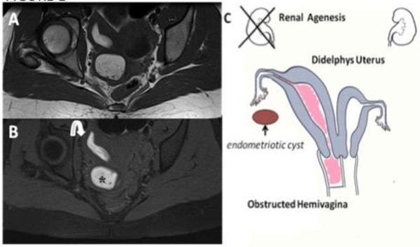
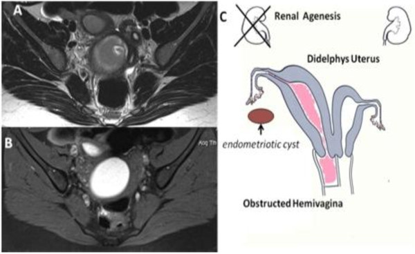
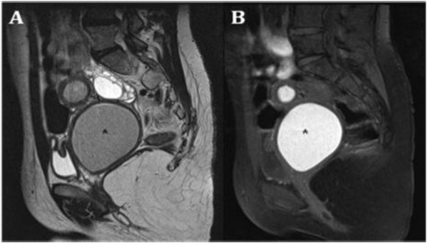
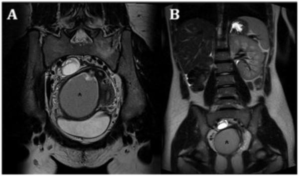
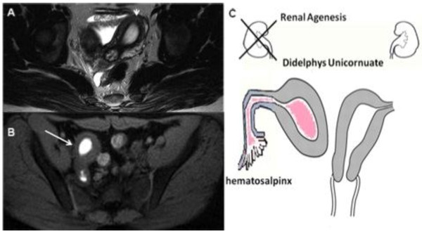
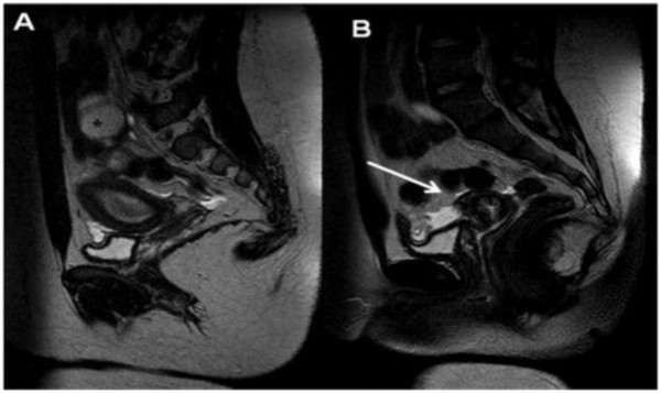
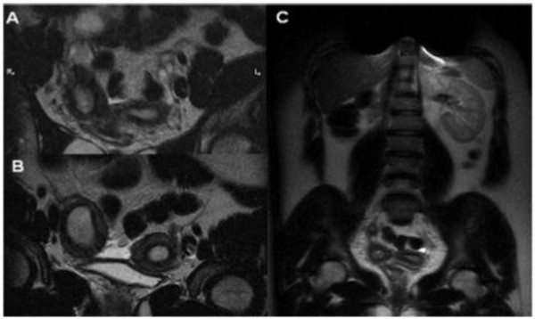
Background: Herlyn-Werner-Wunderlich (HWW) syndrome is a very rare congenital anomaly of the urogenital tract involving Müllerian ducts and Wolffian structures, and it is characterized by the triad of didelphys uterus, obstructed hemivagina and ipsilateral renal agenesis. It generally occurs at puberty and exhibits non-specific and variable symptoms with acute or pelvic pain shortly following menarche, causing a delay in the diagnosis. Moreover, the diagnosis is complicated by the infrequency of this syndrome, because Müllerian duct anomalies (MDA) are infrequently encountered in a routine clinical setting.

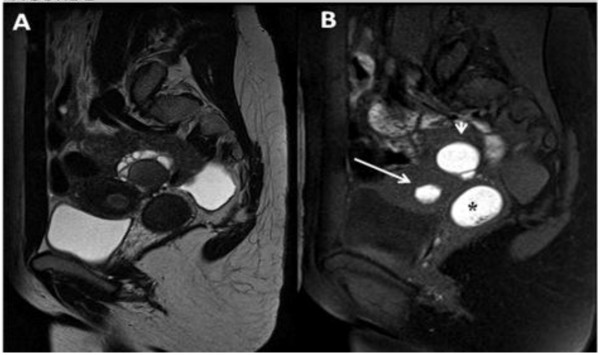
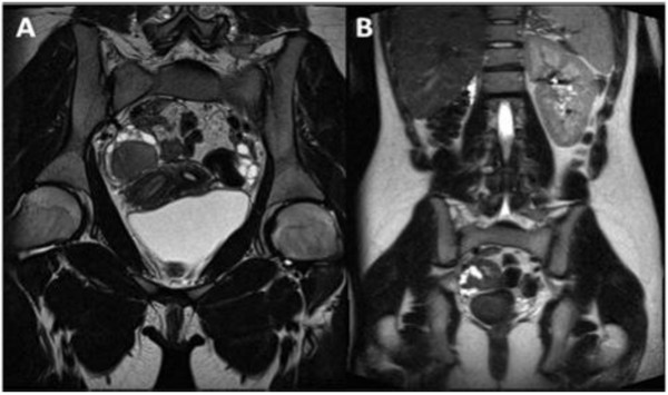
Cases presentation: two cases of HWW syndrome in adolescents and a differential diagnosis for one case of a different MDA, and the impact of magnetic resonance (MR) imaging technology to achieve the correct diagnosis.

Conclusions: MR imaging is a very suitable diagnostic tool in order to perform the correct diagnosis of HWW syndrome.


Figures

Figure 1
Figure 1
Herlyn-Werner-Wunderlich syndrome (C) Drawing illustrates the triad (renal agenesis, didelphys uterus, and obstructed hemivagina and in this case the presence of an endometriosic cyst. A 16-years-old girl presenting the triad of didelphys uterus (class III MDA), an obstructed right hemivagina (class I MDA), and ipsilateral renal agenesis. (A) Axial turbo spin-echo T2 weighted (B) Axial turbo spin-echo T1 fat-saturated weighted MR image showing centrally a hematocolpos (asterisk), a finding corresponding to the obstructed right hemivagina. Mild dilation of the right endometrial cavity (curved arrow) with fluid exhibiting a signal intensity similar to methaemoglobin due to haematometra.
Figure 2
Figure 2
A) Sagittal T2 TSE weighted image showing a mild fluid in the pouch of Douglas. (B) Sagittal T1 TSE fat-suppressed weighted image showing distention of the right endometrial cavity (long straight arrow) with high-signal-intensity material distending the right endometrial cavity and cervix (asterisk), a finding indicative of right hematometrocolpos; on the right ovary presence of an endometriosic cysts appearing hyperintense on T1 fat-saturated image (short straight arrow).
Figure 3
Figure 3
A) Oblique coronal TSE T2-weighted MR image shows a complete view of the anatomical defects. B) Coronal T2 Trufi MR image showing the omolateral renal aplasia with respect to the pelvic abnormalities and a solitary left kidney with a mild hypertrophy.
Figure 4
Figure 4
Herlyn-Werner-Wunderlich syndrome (C) Drawing illustrates the triad. A 15-years-old girl presenting the triad of didelphys uterus (class III MDA), an obstructed right hemivagina (class I MDA), and ipsilateral renal agenesis. (A) Axial turbo spin-echo T2 weighted MR image (B) Axial turbo spin-echo T1 fat-saturated weighted MR image showing centrally a wide hematocolpos (asterisk), corresponding to the obstructed right hemivagina. On the left side the uterus normally communicating with non-obstructed hemivagina.
Figure 5
Figure 5
A) Sagittal T2-weighted MR image demonstrates an enormously dilated hemivagina (asterisk) due to the presence of the transverse septum causing the obstruction and low T2-signal-intensity contents corresponding to a high T1-signal intensity; B) consistent with blood product due to the presence of haemoglobin.
Figure 6
Figure 6
A) Coronal T2 weighted images show the wide hematocolpos (asterisk). B) Trufi coronal image demonstrates the absence of right kidney, omolateral to the pelvic alteration, and a mild compensatory hypertrophy of the left kidney.
Figure 7
Figure 7
A) TSE axial T2 weighted MR image. B) Axial fat-saturated T1-weighted images. C) Drawing illustrates a 14-year-old girl with an unicornuate uterus and rudimentary right horn with an endometrial cavity that does not communicate with the uterus (class II MDA type) and shows an obstructed functional right uterine horn (long straight arrow) containing blood degradation products and a corresponding high T1fs-signal intensity, exhibiting no communication with the cavity of the normal left uterine horn (short straight arrow).
Figure 8
Figure 8
A) Sagittal T2-weighted MR image shows a normal left uterine cavity communicating with a normal cervix (long straight arrow), which in turn communicates with a normal vagina. (B) Right hematosalpinx (asterisk) with a distended non-communicating horn filled with fluid moderately hyperintense on T2 sequences due to haemoglobin degradation products.
Figure 9
Figure 9
A-B-C) Coronal T2 weighted images demonstrate the absence of right kidney, omolateral to the pelvic alteration.

References

    1. Gholoum S, Puligandla PS, Hui T, Su W, Quiros E, Laberge JM. Management and outcome of patients with combined vaginal septum, bifid uterus and ipsilateral renal agenesis (Herlin-Werner-Wunderlich syndrome. J Pediatr Surg. 2006;41(5):987–92. doi: 10.1016/j.jpedsurg.2006.01.021. - DOI - PubMed
    1. Alan JW, Louis RK. Campbell-Walsh Urology. 9. Philadelphia: Saunders; pp. 3270–6.
    1. Orazi C, Lucchetti MC, Schingo PM, Marchetti P, Ferro F. Herlyn-Werner-Wunderlich syndrome: uterus didelphys, blind hemivagina and ipsilateral renal agenesis. Sonographic and MR findings in 11 cases. Pediatr Radiol. 2007;37:657–65. doi: 10.1007/s00247-007-0497-y. - DOI - PubMed
    1. Lee BH, Kim JW, Oh SI. et al.3 cases of uterus didelphys with obstructed hemivagina and ipsilateral renal agenesis. Korean J Obstet Gynecol. 1997;40:1489–95.
    1. Heinonen PK. Clinical implications of the didelphic uterus: long-term follow-up of 49 cases. Eur J Obstet Gynecol Reprod Biol. 2000;91:183–90. doi: 10.1016/S0301-2115(99)00259-6. - DOI - PubMed