. 2012 Apr 10;22(7):638-44.
doi: 10.1016/j.cub.2012.02.030. Epub 2012 Mar 8.

Checkpoint-independent stabilization of kinetochore-microtubule attachments by Mad2 in human cells

Affiliations

Checkpoint-independent stabilization of kinetochore-microtubule attachments by Mad2 in human cells

Lilian Kabeche et al. Curr Biol. .

Abstract

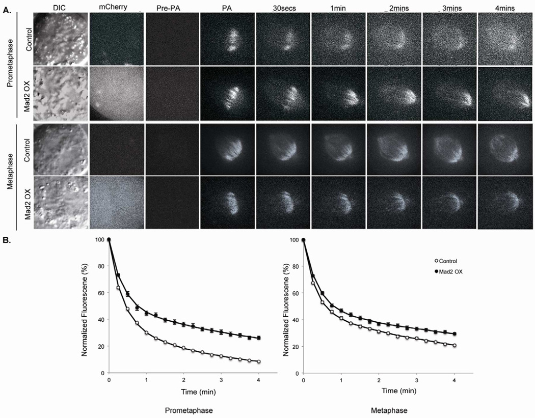
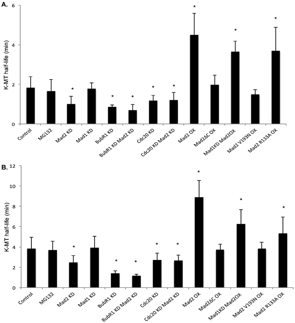
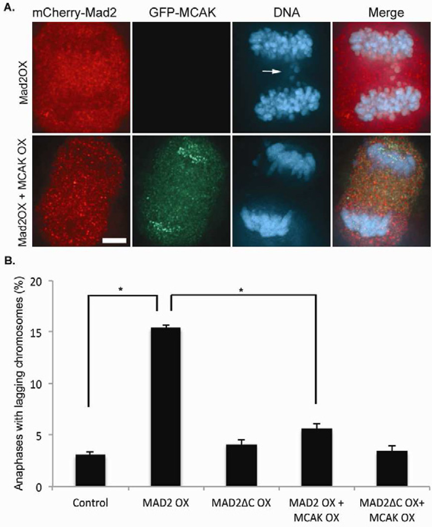
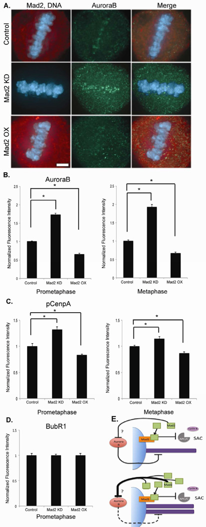
Faithful chromosome segregation is required for cell and organism viability and relies on both the mitotic checkpoint and the machinery that corrects kinetochore-microtubule (k-MT) attachment errors. Most solid tumors have aneuploid karyotypes and many missegregate chromosomes at high rates in a phenomenon called chromosomal instability (CIN). Mad2 is essential for mitotic checkpoint function and is frequently overexpressed in human tumors that are CIN. For unknown reasons, cells overexpressing Mad2 display high rates of lagging chromosomes. Here, we explore this phenomenon and show that k-MT attachments are hyperstabilized by Mad2 overexpression and that this undermines the efficiency of correction of k-MT attachment errors. Mad2 affects k-MT attachment stability independently of the mitotic checkpoint because k-MT attachments are unaltered upon Mad1 depletion and Mad2 overexpression hyperstabilizes k-MT attachments in Mad1-deficient cells. Mad2 mediates these effects with Cdc20 by altering the centromeric localization and activity of Aurora B kinase, a known regulator of k-MT attachment stability. These data reveal a new function for Mad2 to stabilize k-MT attachments independent of the checkpoint and explain why Mad2 overexpression increases chromosome missegregation to cause chromosomal instability in human tumors.


Figures

Figure 1
Figure 1
Measurement of kinetochore-microtubule dynamics in cells overexpressing Mad2. (A) DIC and time-lapse fluorescent images of prometaphase and metaphase spindles in untreated (control) and Mad2 overexpression (Mad2OX) in PA-GFP tubulin expressing RPE1 cells before (Pre-PA) and at indicated times after photoactivation. Mad2 overexpression is visualized by mCherry fluorescence. (B) Normalized fluorescence intensity over time after photoactivation of spindles in untreated (white circles) and Mad2OX (filled circles) in prometaphase and metaphase cells. Data represents mean ± s.e.m., n≥10 cells.
Figure 2
Figure 2
Kinetochore-microtubule half-life. kMT half-life calculated from the exponential decay curve of photoactivated fluorescence (r2 > 0.99) under different conditions for (A) prometaphase and (B) metaphase. Error bars represent SD, *p<0.05, t-test, n=10–20 cells.
Figure 3
Figure 3
MCAK overexpression suppresses lagging chromosomes in anaphase in Mad2 overexpressing cells. (A) Anaphase in RPE-1 cells expressing mCherry-Mad2 (red) alone or both mCherry-Mad2 and GFP-MCAK (green) and stained for DNA (blue). The lagging chromosomes induced by Mad2 overexpression are highlighted by an arrow. Scale bar 5µm. (B) Number of anaphases with lagging chromosomes in untreated RPE-1 cells (control) and RPE-1 cells overexpressing mCherry-Mad2 (Mad2OX), mCherry-Mad2ΔC (Mad2ΔCOX), mCherry-Mad2 and GFP-MCAK (Mad2OX + MCAKOX), and mCherry−Mad2ΔC and GFP-MCAK (Mad2ΔCOX + MCAKOX). Bars represent mean ± s.e.m., *p<0.05, t-test, n=300 cells of three experiments.
Figure 4
Figure 4
Mad2 affects Aurora B localization and activity at the centromeres. (A) Metaphase of untreated RPE-1 cells (control) and RPE-1 cells overexpressing Mad2 (Mad2OX) and depleted of Mad2 (Mad2KD). Scale bar 5µm. Fluorescence intensities of centromeres stained for (B) Aurora B and (C) phosphorylated CenpA (pCenpA) in RPE1 cells normalized using CREST in both prometaphase and metaphase. (D) Fluorescence intensity of BubR1 at kinetochores in prometaphase. Bars represent mean ± s.e.m., *p<0.05, t-test, n=150–200 kinetochore pairs (15–20 cells) of three experiments. (E) Model for Mad2 stabilization of kinetochore-microtubules. Mad2 promotes k-MT stability by attenuating the destabilizing effect of Aurora B activity on k-MT attachments. The question mark refers to intermediate components of this pathway that lie between Mad2 and Aurora B that are yet to be identified. Overexpression of Mad2 excessively suppresses Aurora B activity resulting in hyperstable k-MT attachments.

Comment in

References

    1. Musacchio A, Salmon ED. The spindle-assembly checkpoint in space and time. Nat. Rev. Mol. Cell Biol. 2007;8:379–393. - PubMed
    1. Cimini D, Howell B, Maddox P, Khodjakov A, Degrassi F, Salmon ED. Merotelic kinetochore orientation is a major mechanism of aneuploidy in mitotic mammalian tissue cells. J Cell Biol. 2001;153:517–527. - PMC - PubMed
    1. Bakhoum SF, Thompson SL, Manning AL, Compton DA. Genome stability is ensured by temporal control of kinetochore-microtubule dynamics. Nat. Cell. Biol. 2009;11:27–35. - PMC - PubMed
    1. Lengauer C, Kinzler KW, Vogelstein B. Genetic instabilities in human cancers. Nature. 1998;396:643–649. - PubMed
    1. Thompson SL, Compton DA. Examining the link between chromosomal instability and aneuploidy in human cells. J. Cell. Biol. 2008;180:665–672. - PMC - PubMed

Publication types

MeSH terms