. 2012 Apr 17;22(4):749-62.
doi: 10.1016/j.devcel.2012.02.001. Epub 2012 Mar 8.

HJURP uses distinct CENP-A surfaces to recognize and to stabilize CENP-A/histone H4 for centromere assembly

Affiliations

HJURP uses distinct CENP-A surfaces to recognize and to stabilize CENP-A/histone H4 for centromere assembly

Emily A Bassett et al. Dev Cell. .

Abstract

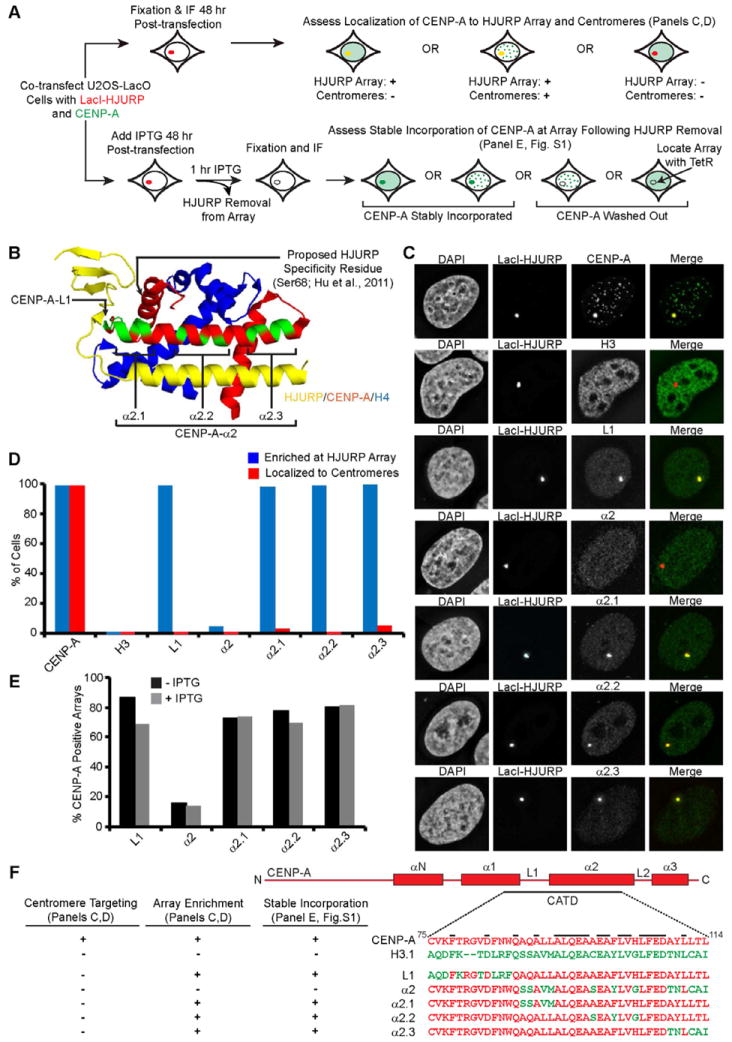
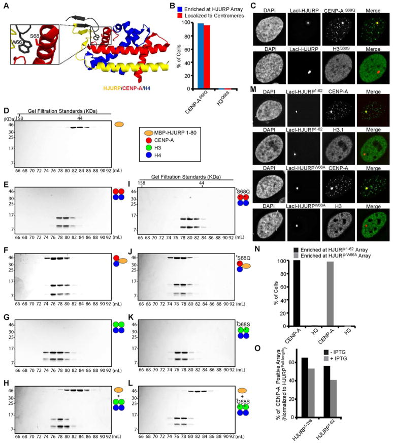
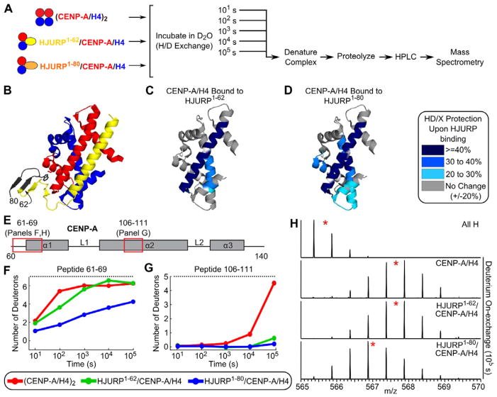
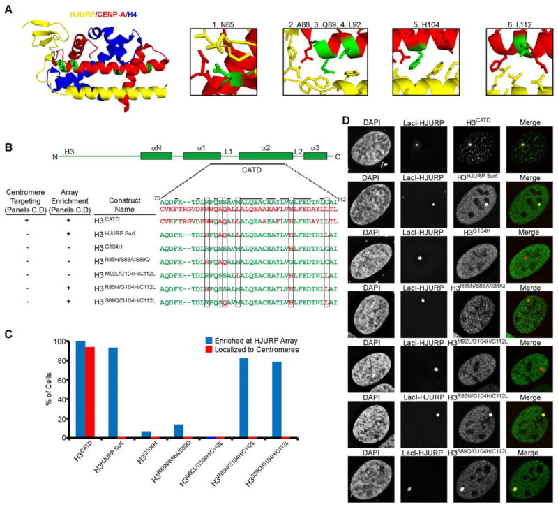
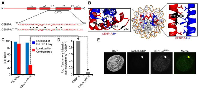
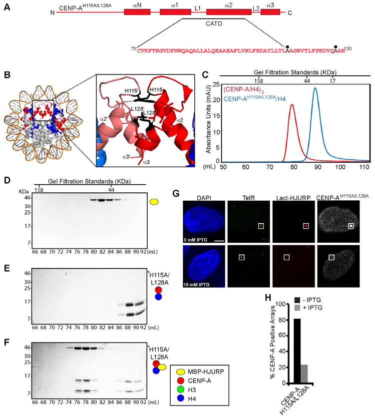
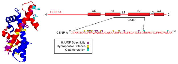
Centromeres are defined by the presence of chromatin containing the histone H3 variant, CENP-A, whose assembly into nucleosomes requires the chromatin assembly factor HJURP. We find that whereas surface-exposed residues in the CENP-A targeting domain (CATD) are the primary sequence determinants for HJURP recognition, buried CATD residues that generate rigidity with H4 are also required for efficient incorporation into centromeres. HJURP contact points adjacent to the CATD on the CENP-A surface are not used for binding specificity but rather to transmit stability broadly throughout the histone fold domains of both CENP-A and H4. Furthermore, an intact CENP-A/CENP-A interface is a requirement for stable chromatin incorporation immediately upon HJURP-mediated assembly. These data offer insight into the mechanism by which HJURP discriminates CENP-A from bulk histone complexes and chaperones CENP-A/H4 for a substantial portion of the cell cycle prior to mediating chromatin assembly at the centromere.


Figures

Figure 1
Figure 1. HJURP binding is insufficient for centromere targeting of CENP-A
(A) Scheme for HJURP chromosome tethering assay in U2OS-LacO-TRE cells. In this system, the mCherry-LacI-HJURP fusion protein is expressed at similar levels to endogenous HJURP (Fig. S1B) (B) Structure of HJURP/CENP-A/H4 complex (PDB 3R45; Hu et al., 2011) highlighting CATD residues swapped for H3 (green) in the various mutant versions. The location of the proposed HJURP specificity determinant (Ser68; Hu et al., 2011) is also shown. (C) Representative images of CENP-A, H3, or mutant versions of CENP-A introduced along with LacI-HJURP into U2OS-LacO-TRE cells. (D) Quantification of the sub-nuclear localization of the indicated histone constructs. In each case, 100 cells were counted and the results are representative of multiple independent experiments. (E) Quantification of stable CENP-A incorporation into the HJURP-containing array. Cells were analyzed 48 h after co-transfection of mCherry-LacI-HJURP, GFP-TetR, and HA-tagged CENP-A mutant proteins and a 1 h treatment with (gray) or without (black) 15 mM IPTG. (F) Sequences of the CENP-A mutants used in these experiments and a summary of our results. Black bars indicate residues shared in both H3 and CENP-A. See also Figure S1.
Figure 2
Figure 2. CENP-A Ser68 is not a recognition determinant for HJURP binding
(A) Highlight of the contact point between CENP-A Ser68 and HJURP Trp66 (PDB 3R45; Hu et al., 2011). The region of HJURP deleted in the HJURP1–62 version is labeled in black. (B) Quantification of the sub-nuclear localization of the indicated mutant histone constructs. WT histones were tested in parallel and those data are shown in Fig. S2M,O. (C) Representative images of the indicated mutant histone constructs introduced along with LacI-HJURP into U2OS-LacI-TRE cells. (D–L) SDS-PAGE of the indicated fractions from SEC of the indicated protein mixes. (M) mCherry-LacI-HJURP1–62 or full length mCherry-LacI-HJURPW66A was co-transfected into U2OS-LacO-TRE cells with HA-CENP-A or HA-H3 and cells were analyzed at 48 hours. WT histones were tested in parallel and those data are shown in Fig. S2N,P. (N) Quantification of CENP-A and H3 recruitment to HJURP1–62 and HJURPW66A arrays. (O) Quantification of stable incorporation of endogenous CENP-A into the HJURP-containing array. Cells co-transfected with mCherry-LacI-HJURP1–62 and GFP-TetR were treated 48 h post-transfection with (gray) or without (black) 15 mM IPTG for 1 h, and assessed for recruitment of endogenous CENP-A to the array. The values shown are normalized to the level of CENP-A recruitment to mCherry-LacI-HJURPFull-length. See also Figure S2.
Figure 3
Figure 3. HJURP interactions with the α1 helix of CENP-A stabilize the histone fold domains of both CENP-A and H4
(A) Experimental scheme for determining H/DX of protein complexes at various time points. (B) Structure of HJURP/CENP-A/H4 complex (PDB 3R45; Hu et al., 2011). The region of HJURP absent from the HJURP1–62 version is labeled in black. (C–D) Protection from H/DX upon binding HJURP1–62 (C) or HJURP1–80 (D) is mapped onto the structure of CENP-A/H4 (PDB 3NQJ; Sekulic et al., 2010). These data correspond to the 105 s time point. Labeling is as indicated in the legend, indicating the consensus behavior of all overlapping peptides at each position (white indicates the small number of positions lacking peptide coverage). (E) Diagram of CENP-A secondary structure with red boxes corresponding to locations of example peptides shown in F–G. (F–G) Comparison of H/DX for the indicated CENP-A peptides from each of the indicated complexes. Dotted lines indicate maximum levels of deuteration determined from fully-deuterated samples. (H) Raw mass spectrometry data for the CENP-A peptide shown in (F). Red star indicates peptide centroid value. ‘All H’ is data from the non-deuterated control sample. See also Figure S3.
Figure 4
Figure 4. As few as three residues within the CATD are sufficient for HJURP specificity
(A) Structure of HJURP/CENP-A/H4 structure (PDB 3R45; Hu et al., 2011) highlighting six candidate residues within the CATD to confer HJURP specificity. (B) Sequences of versions of H3 carrying the indicated CATD residues and summary of our results. Black bars indicate residues shared in both H3 and CENP-A, and boxes highlight the six candidate residues. (C) Quantification of the sub-nuclear localization of the indicated histone constructs. In each case, 100 cells were counted and the results are representative of multiple independent experiments. (D) Representative images of the indicated histone constructs introduced along with mCherry-LacI-HJURP into U2OS-LacO-TRE cells. See also Figure S4.
Figure 5
Figure 5. CENP-A hydrophobic stitch residues are required for assembly into centromeric chromatin
(A) Sequence of the CENP-AH4 Int. mutant containing six H3 residues, highlighted with black circles. (B) Structure of the CENP-A-containing nucleosome (PDB 3AN2; Tachiwana et al., 2011) highlighting the six CENP-A a.a. positions substituted with H3 residues in CENP-AH4 Int.. (C) Quantification of the sub-nuclear localization of CENP-AH4 Int.. (D) The average centromere intensity was measured for the population of cells in (C) with indicated histone localizing to the centromere. Intensity is normalized to wild-type CENP-A. (E) Representative images of CENP-AH4 Int. introduced along with LacI-HJURP into U2OS-LacO-TRE cells. See also Figure S5.
Figure 6
Figure 6. An intact CENP-A/CENP-A interface is a requirement for HJURP-mediated nucleosome assembly
(A) Diagram of the CENP-AH115A/L128A mutant protein. Black circles indicate mutated residues. (B) Structure of the CENP-A nucleosome (PDB 3AN2; Tachiwana et al., 2011), with inset depicting residues H115 and L128 within the CENP-A/CENP-A interface. (C) Gel filtration chromatograph comparing elution profiles of (CENP-A/H4)2 and (CENP-AH115A/L128A/H4). (D–F) SDS-PAGE of the indicated fractions from SEC of the indicated protein mixes. (G) Representative images of cells co-transfected with mCherry-LacI-HJURP, GFP-TetR, and HA-tagged CENP-AH115A/L128A and treated with or without IPTG for 1 h prior to processing for immunofluorescence. (H) Quantification of CENP-AH115A/L128A stable incorporation into the HJURP-containing array with (gray) or without (black) 15 mM IPTG. At least 30 cells were counted over multiple experiments. See also Figure S6.
Figure 7
Figure 7. Summary of CENP-A features required for centromeric nucleosome assembly
Structure of CENP-A/H4 (PDB 3NQJ; Sekulic et al., 2011) highlighting CENP-A residues required for HJURP specificity (purple), a rigid interface with H4 (yellow), and formation of an intact CENP-A/CENP-A interface required for assembling into octameric nucleosomes (cyan).

References

    1. Allshire RC, Karpen GH. Epigenetic regulation of centromeric chromatin: old dogs, new tricks? Nat Rev Genet. 2008;9:923–937. - PMC - PubMed
    1. Amor DJ, Bentley K, Ryan J, Perry J, Wong L, Slater H, Choo KH. Human centromere repositioning “in progress”. Proc Natl Acad Sci U S A. 2004;101:6542–6547. - PMC - PubMed
    1. Barnhart MC, Kuich PH, Stellfox ME, Ward JA, Bassett EA, Black BE, Foltz DR. HJURP is a CENP-A chromatin assembly factor sufficient to form a functional de novo kinetochore. J Cell Biol. 2011;194:229–243. - PMC - PubMed
    1. Bassett EA, Wood S, Salimian KJ, Ajith S, Foltz DR, Black BE. Epigenetic centromere specification directs aurora B accumulation but is insufficient to efficiently correct mitotic errors. J Cell Biol. 2010;190:177–185. - PMC - PubMed
    1. Belmont AS. Visualizing chromosome dynamics with GFP. Trends Cell Biol. 2001;11:250–257. - PubMed

Publication types

MeSH terms

LinkOut - more resources