

Remodeling of mechanical junctions and of microtubule-associated proteins accompany cardiac connexin43 lateralization
- PMID: 22406144
- PMCID: PMC3723688
- DOI: 10.1016/j.hrthm.2012.03.003
Remodeling of mechanical junctions and of microtubule-associated proteins accompany cardiac connexin43 lateralization
Abstract
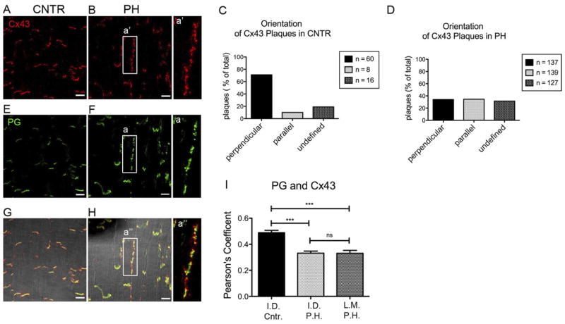
Background: Desmosomes and adherens junctions provide mechanical continuity between cardiac cells, whereas gap junctions allow for cell-cell electrical/metabolic coupling. These structures reside at the cardiac intercalated disc (ID). Also at the ID is the voltage-gated sodium channel (VGSC) complex. Functional interactions between desmosomes, gap junctions, and VGSC have been demonstrated. Separate studies show, under various conditions, reduced presence of gap junctions at the ID and redistribution of connexin43 (Cx43) to plaques oriented parallel to fiber direction (gap junction "lateralization").
Objective: To determine the mechanisms of Cx43 lateralization, and the fate of desmosomal and sodium channel molecules in the setting of Cx43 remodeling.
Methods: Adult sheep were subjected to right ventricular pressure overload (pulmonary hypertension). Tissue was analyzed by quantitative confocal microscopy and by transmission electron microscopy. Ionic currents were measured using conventional patch clamp.
Result: Quantitative confocal microscopy demonstrated lateralization of immunoreactive junctional molecules. Desmosomes and gap junctions in lateral membranes were demonstrable by electron microscopy. Cx43/desmosomal remodeling was accompanied by lateralization of 2 microtubule-associated proteins relevant for Cx43 trafficking: EB1 and kinesin protein Kif5b. In contrast, molecules of the VGSC failed to reorganize in plaques discernable by confocal microscopy. Patch-clamp studies demonstrated change in amplitude and kinetics of sodium current and a small reduction in electrical coupling between cells.
Conclusions: Cx43 lateralization is part of a complex remodeling that includes mechanical and gap junctions but may exclude components of the VGSC. We speculate that lateralization results from redirectionality of microtubule-mediated forward trafficking. Remodeling of junctional complexes may preserve electrical synchrony under conditions that disrupt ID integrity.
Copyright © 2012 Heart Rhythm Society. Published by Elsevier Inc. All rights reserved.
Figures


Comment in
-
Lateralized gap junctions in pulmonary hypertension: lost but not alone.Heart Rhythm. 2012 Jul;9(7):1141-2. doi: 10.1016/j.hrthm.2012.03.053. Epub 2012 Mar 25. Heart Rhythm. 2012. PMID: 22452795 No abstract available.
Similar articles
-
The organization of adherens junctions and desmosomes at the cardiac intercalated disc is independent of gap junctions.J Cell Sci. 2003 Mar 1;116(Pt 5):875-85. doi: 10.1242/jcs.00258. J Cell Sci. 2003. PMID: 12571285
-
Connexin43 regulates sodium current; ankyrin-G modulates gap junctions: the intercalated disc exchanger.Cardiovasc Res. 2012 Feb 1;93(2):220-2. doi: 10.1093/cvr/cvr343. Epub 2011 Dec 16. Cardiovasc Res. 2012. PMID: 22180603
-
Silencing of desmoplakin decreases connexin43/Nav1.5 expression and sodium current in HL‑1 cardiomyocytes.Mol Med Rep. 2013 Sep;8(3):780-6. doi: 10.3892/mmr.2013.1594. Epub 2013 Jul 18. Mol Med Rep. 2013. PMID: 23877755
-
[Remodeling of cardiac gap junctions and arrhythmias].Sheng Li Xue Bao. 2011 Dec 25;63(6):586-92. Sheng Li Xue Bao. 2011. PMID: 22193455 Review. Chinese.
-
The cardiac connexome: Non-canonical functions of connexin43 and their role in cardiac arrhythmias.Semin Cell Dev Biol. 2016 Feb;50:13-21. doi: 10.1016/j.semcdb.2015.12.002. Epub 2015 Dec 7. Semin Cell Dev Biol. 2016. PMID: 26673388 Free PMC article. Review.
Cited by
-
Apelin Ameliorates High Glucose-Induced Downregulation of Connexin 43 via AMPK-Dependent Pathway in Neonatal Rat Cardiomyocytes.Aging Dis. 2018 Feb 1;9(1):66-76. doi: 10.14336/AD.2017.0426. eCollection 2018 Feb. Aging Dis. 2018. PMID: 29392082 Free PMC article.
-
Desmosomal Junctions and Connexin-43 Remodeling in High-Pacing-Induced Heart Failure Dogs.Anatol J Cardiol. 2023 Aug 1;27(8):462-471. doi: 10.14744/AnatolJCardiol.2023.2823. Epub 2023 Jun 7. Anatol J Cardiol. 2023. PMID: 37288855 Free PMC article.
-
A microtubule-connexin-43 regulatory link suppresses arrhythmias and cardiac fibrosis in Duchenne muscular dystrophy mice.Am J Physiol Heart Circ Physiol. 2022 Nov 1;323(5):H983-H995. doi: 10.1152/ajpheart.00179.2022. Epub 2022 Oct 7. Am J Physiol Heart Circ Physiol. 2022. PMID: 36206047 Free PMC article.
-
Desmosomal junctions are necessary for adult sinus node function.Cardiovasc Res. 2016 Aug 1;111(3):274-86. doi: 10.1093/cvr/cvw083. Epub 2016 Apr 20. Cardiovasc Res. 2016. PMID: 27097650 Free PMC article.
-
Arrhythmogenic cardiomyopathy and Brugada syndrome: diseases of the connexome.FEBS Lett. 2014 Apr 17;588(8):1322-30. doi: 10.1016/j.febslet.2014.02.008. Epub 2014 Feb 15. FEBS Lett. 2014. PMID: 24548564 Free PMC article. Review.
References
-
- Tan XY, He JG. The remodeling of connexin in the hypertrophied right ventricular in pulmonary arterial hypertension and the effect of a dual ET receptor antagonist (bosentan) Pathol Res Pract. 2009;205:473–482. - PubMed
Publication types
MeSH terms
Substances
Grants and funding
LinkOut - more resources
Full Text Sources
Medical
Miscellaneous
