Review
. 2012 Oct;8(7):1052-69.
doi: 10.1016/j.nano.2012.02.009. Epub 2012 Mar 7.

NTS-Polyplex: a potential nanocarrier for neurotrophic therapy of Parkinson's disease

Affiliations
Review

NTS-Polyplex: a potential nanocarrier for neurotrophic therapy of Parkinson's disease

Daniel Martinez-Fong et al. Nanomedicine. 2012 Oct.

Abstract

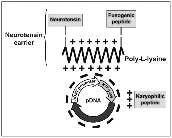
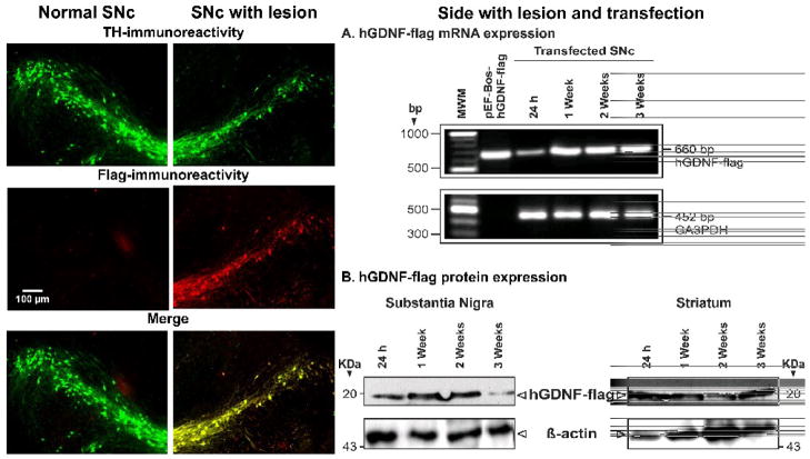
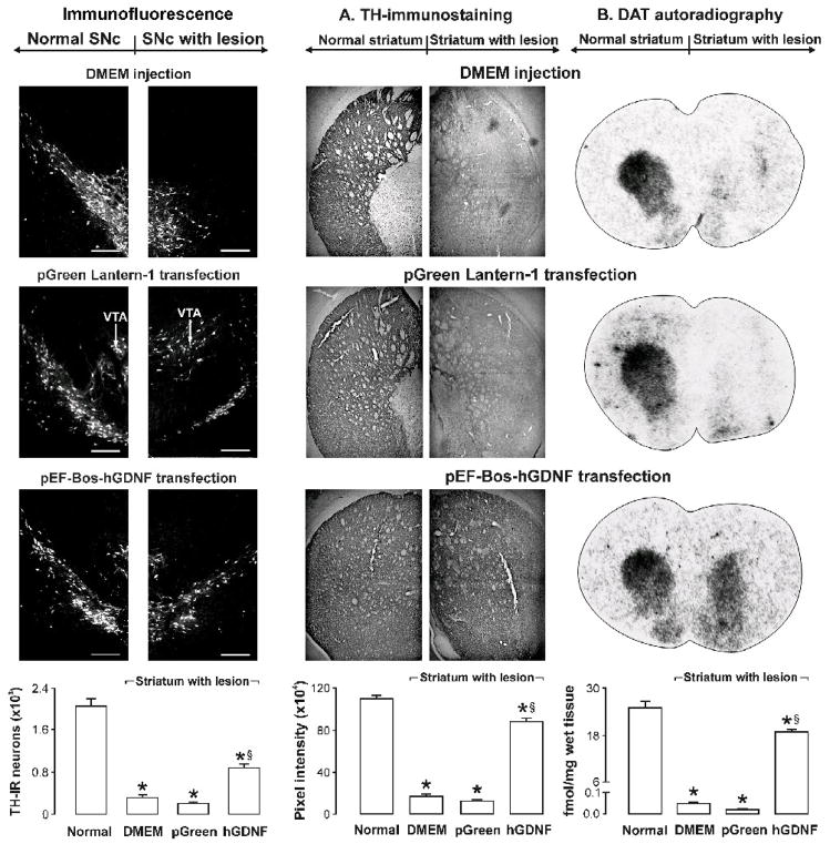
Nanomedicine has focused on targeted neurotrophic gene delivery to the brain as a strategy to stop and reverse neurodegeneration in Parkinson's disease. Because of improved transfection ability, synthetic nanocarriers have become candidates for neurotrophic therapy. Neurotensin (NTS)-polyplex is a "Trojan horse" synthetic nanocarrier system that enters dopaminergic neurons through NTS receptor internalization to deliver a genetic cargo. The success of preclinical studies with different neurotrophic genes supports the possibility of using NTS-polyplex in nanomedicine. In this review, we describe the mechanism of NTS-polyplex transfection. We discuss the concept that an effective neurotrophic therapy requires a simultaneous effect on the axon terminals and soma of the remaining dopaminergic neurons. We also discuss the future of this strategy for the treatment of Parkinson's disease.

From the clinical editor: This review paper focuses on nanomedicine-based treatment of Parkinson's disease, a neurodegenerative condition with existing symptomatic but no curative treatment. Neurotensin-polyplex is a synthetic nanocarrier system that enables delivery of genetic cargo to dopaminergic neurons via NTS receptor internalization.


Figures

Figure 1
Figure 1
Diagram showing that the branching of dopaminergic axons originating in the SNc is quite extensive, innervating several nuclei of the brain. The lesion of dopaminergic neuron cell bodies can thus reduce dopamine transmission in multiple structures, thus causing the motor and nonmotor manifestations of Parkinson’s disease.
Figure 2
Figure 2
Ability of NTS-polyplex to transfect dopaminergic neurons in vitro and in vivo. Top panel. Confocal micrographs showing the GFP expression in cultured dopaminergic neurons after transfection with the plasmid pEGFP-N1. NTS-polyplex was formed at 1:833:36 molar ratio (pEGFP-N1, 6 nM: PK, 5 μM: NTS-vector, 216 nM) in a mixture of Neurobasal medium and B27, serum free, and applied immediately to 2-day-old primary cultures. After 6-h exposure, the transfection medium was replaced by fresh culture medium and cells were incubated for an additional 72 h, as described previously. Cells were subjected to double immunofluorescence against GFP and TH. The primary antibodies were a rabbit polyclonal antibody to GFP (Abcam; Cambridge MA, USA) and a mouse monoclonal antibody to TH (Sigma-Aldrich, St. Louis, MO, USA). The secondary antibodies were an Alexa 488 chicken anti-rabbit (Molecular Probes Inc., Eugene, OR, USA) and a donkey anti-mouse TRITC (Jackson Immunoresearch Laboratories Inc., West Grove, PA, USA). Calibration bars = 50 μm. Bottom panel. Confocal micrographs showing the GFP expression in dopaminergic neurons of the substantia nigra after the local injection of the NTS-polyplex with pDAT-EGFP. Calibration bars = 200 μm. Micrographs of panel B were reproduced from Arango-Rodriguez et al. (2006).
Figure 3
Figure 3
Schematic illustration of the sequence of NTS-polyplex-mediated gene transfection documented by confocal microscopy in neuroblastoma N1E-115 cells. A. NTS-polyplex harboring a propidium iodide-labeled plasmid DNA was used for transfection and calcein was used to delimit the cell area. The micrographs of the first row were taken 5 min after incubation with propidium iodide-labeled NTS-polyplex in the presence of 0.45 M sucrose to block receptor-mediated endocytosis . The incubation time with 0.45 M sucrose was 30 min.,, B. GFP expression in N1E-115 cells counterstained with Hoechst, a nuclear staining. The microphotographs were taken at the times after transfection shown at the left margins. Calibration bars = 20 μm. The transfection efficiency is shown in figures 6 and 7 (in vitro) and figures 2 and 9 (in vivo). The first and third panels were reproduced from Navarro-Quiroga et al. (2002).
Figure 4
Figure 4
Electron microscopy analysis of the sequential steps of the formation of the NTS-polyplex nanoparticles with pEGFP-N1. The micrographs show the natural form of 6 nM pDNA alone (A), the initial condensation of pDNA caused by the addition of 6 μM KP (B), the condensation of the KP-pDNA complex in the presence of 1% FBS (C), and the toroidal condensation of the KP-pDNA complex caused by the addition of the NTS-vector at the optimum molar ratio (1:34) to form the NTS-polyplex nanoparticles (D). Micrographs were reproduced from Arango-Rodriguez et al. (2006).
Figure 5
Figure 5
Scheme of the NTS-polyplex components. The neurotensin carrier is the conjugate of neurotensin and fusogenic peptide with poly-L-lysine. The karyophilic peptide is electrostatically bound to a plasmid DNA (pDNA) to form the KP-pDNA complex, which is bound to the neurotensin carrier to form the NTS-polyplex. The pDNA encompasses the gene of interest under control of a tissue-specific promoter; for instance, hDAT (human dopamine transporter), a promoter specific for dopaminergic neurons. NTS = pyroGlu-Leu-Tyr-Glu-Asn-Lys-Pro-Arg-Arg-Pro-Tyr-Ile-Leu-OH. Modified hemagglutinin HA2 FP = Gly-Leu-Phe-Glu-Ala-Ile-Ala-Glu-Phe-Ile-Glu-Gly-Gly-Trp-Glu-Gly-Leu-Ile-Glu-Gly-Cys-Ala-Lys-Lys-Lys-OH. SV40 Vp1 NLS = Met-Ala-Pro-Thr-Lys-Arg-Lys-Gly-Ser-Cys-Pro-Gly-Ala-Ala-Pro-Asn-Lys-Pro-Lys-OH.
Figure 6
Figure 6
Flow cytometry analysis showing the contribution of fusogenic and karyophilic peptides to the improvement of NTS-polyplex internalization. The plasmid was labeled with propidium iodide. (A) Basal fluorescence in N1E-115 cells.(B) Blockade by 100 nM SR-48692 of fusogenic-karyophilic-NTS-polyplex internalization in N1E-115 cells. (C) Failure of fusogenic-karyophilic-NTS-polyplex to internalize in COS7 cells, which lack NTSR1. (D) Internalization of karyophilic-NTS-polyplex in N1E-115 cells.(E) Internalization of fusogenic-NTS-polyplex in N1E-115 cells.(F) Internalization of fusogenic-karyophilic-NTS-polyplex in N1E-115 cells. Populations of 104 cells emitting red fluorescence of propidium iodide were distributed in three arbitrary regions (R1, R2 and R3) according to their fluorescence intensity. *, Significantly different from R3 in (D); **, significantly different from R3 in (E). The figure was reproduced from Navarro-Quiroga et al. (2002).
Figure 7
Figure 7
Flow cytometry analysis showing the contribution of fusogenic and karyophilic peptides to the improvement of NTS-polyplex-mediated gene expression. pGreen Lantern 1 was delivered by different polypplexes. (A) Basal fluorescence in N1E-115 cells. (B) Lack of GFP expression in N1E-115 cells exposed to the fusogenic-karyophilic-NTS-polyplex in the presence of 100 nM SR-48692. (C) Lack of GFP expression in COS7 exposed to fusogenic-karyophilic-NTS-polyplex. (D) GFP expression in N1E-115 cells transfected by karyophilic-NTS-polyplex. (E) GFP expression in N1E-115 cells transfected by karyophilic-NTS-polyplex fusogenic-NTS-polyplex. (F) GFP expression in N1E-115 cells transfected by karyophilic-NTS-polyplex fusogenic-karyophilic-NTS-polyplex. Populations of 104 cells emitting the GFP fluorescence were distributed in three arbitrary regions (R1, R2 and R3) according to their fluorescence intensity. *, Significantly different from R3 in (D); **, significantly different from R3 in (E). The figure was reproduced from Navarro-Quiroga et al. (2002).
Figure 8
Figure 8
Sites of the 6-OHDA lesion and NTS-polyplex transfection. 6-OHDA was injected in the striatum at the coordinates: anteroposterior (AP) +7.7 mm from interaural line; mediolateral (ML) + 4.0 mm from interparietal suture and dorsoventral (DV) −5.4 mm from dura mater. One or twelve weeks after 6-OHDA injection, different NTS-polyplexes were injected into the ipsilateral substantia nigra at the coordinates AP + 2.5 mm from interaural line, ML + 2.0 mm from midline and DV −6.7 from dura mater. The coordinates were adapted from the Paxinos atlas for rats weighing 220 g.
Figure 9
Figure 9
The overexpression of hGDFN gene in dopaminergic neurons of the substantia nigra leads to the presence of hGDFN protein in the striatum, suggesting the axonal transport of the transgenic protein to the other nuclei innervated by nigral dopaminergic neurons. The micrographs of double immunofluorescence against TH and flag show hGDNF-flag expression in dopamine neurons of the substantia nigra of hemiparkinsonian rats at 3 weeks following intranigral transfection of pEF-BoshGDNF-flag using NTS-polyplex. The calibration bar in the left middle panel is valid for all other panels. A) The representative RT-PCR assays shows the time course of hGDNF-flag gene expression in the substantia nigra of hemiparkinsonian rats. B) The western blot assay shows the presence of hGDNF-flag protein in the substantia nigra and striatum. The figures were reproduced from Gonzalez-Barrios et al. (2006).
Figure 10
Figure 10
pEF-Bos-hGDNF transfection in the substantia nigra promotes survival in the nigral dopaminergic neurons and reinnervation of the striatum in hemiparkinsonian rats. The figures were reproduced from Gonzalez-Barrios et al. (2006).

References

    1. Galvan A, Wichmann T. Pathophysiology of parkinsonism. Clin Neurophysiol. 2008;119:1459–74. - PMC - PubMed
    1. Kish SJ, Shannak K, Hornykiewicz O. Uneven pattern of dopamine loss in the striatum of patients with idiopathic Parkinson’s disease. Pathophysiologic and clinical implications. N Engl J Med. 1988;318:876–80. - PubMed
    1. Brooks DJ. PET studies on the function of dopamine in health and Parkinson’s disease. Ann N Y Acad Sci. 2003;991:22–35. - PubMed
    1. Chaudhuri KR, Schapira AH. Non-motor symptoms of Parkinson’s disease: dopaminergic pathophysiology and treatment. Lancet Neurol. 2009;8:464–74. - PubMed
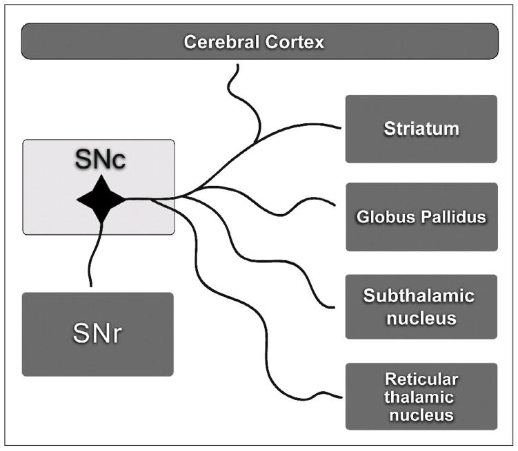
    1. Anaya-Martinez V, Martinez-Marcos A, Martinez-Fong D, Aceves J, Erlij D. Substantia nigra compacta neurons that innervate the reticular thalamic nucleus in the rat also project to striatum or globus pallidus: implications for abnormal motor behavior. Neuroscience. 2006;143:477–86. - PubMed

Publication types