Review
. 2012 Apr;60(4):643-63.
doi: 10.2310/JIM.0b013e31824a4d79.

Pancreatic ductal adenocarcinoma: a review of immunologic aspects

Affiliations
Review

Pancreatic ductal adenocarcinoma: a review of immunologic aspects

Megan B Wachsmann et al. J Investig Med. 2012 Apr.

Abstract

With the continued failures of both early diagnosis and treatment options for pancreatic cancer, it is now time to comprehensively evaluate the role of the immune system on the development and progression of pancreatic cancer. It is important to develop strategies that harness the molecules and cells of the immune system to treat this disease. This review will focus primarily on the role of immune cells in the development and progression of pancreatic ductal adenocarcinoma and to evaluate what is known about the interaction of immune cells with the tumor microenvironment and their role in tumor growth and metastasis. We will conclude with a brief discussion of therapy for pancreatic cancer and the potential role for immunotherapy. We hypothesize that the role of the immune system in tumor development and progression is tissue specific. Our hope is that better understanding of this process will lead to better treatments for this devastating disease.


Figures

Figure 1
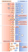
Figure 1. A–B Interaction between PDAC and the Microenvironment
A. The pro- and anti-angiogenic function of Pancreatic Stelate (PS) cells. Immunocytes can release cytokines and growth factors that promote neo angiogenesis as well as activate PS cells. Upon activation by stress, ROS, cytokines and/or growth factors PS cells can secrete periostin to mediate endothelial cell adhesion and migration as well as secrete MMPs. MMPs can both promote neo angiogenesis through basal membrane destruction (MMP-2, MMP-9) or inhibit neo angiogenesis by triggering production of endostatin (MMP-12). Pancreatic cancer cells can also promote neo angiogenesis by stimulating PS cells to secrete VEGF or inhibit neo angiogenesis by increasing endostatin secretion. B. Relationship between insulin resistance and pancreatic cancer development and survival. Insulin resistance can lead to increased insulin and glucose in the blood. When the level of IGF-1 is low in the tumor microenviroment, IGFRs and IRs that can be overexpressed on cancer cells are free for insulin to bind and stimulate cancer cell growth. Furthermore, downstream signaling of the PI3K/Akt/mTOR pathway can sustain cellular survival through the synthesis of anti-apoptotic proteins. Under hypoxic conditions, insulin can mediate VEGF secretion from pancreatic cancer cells via expression of HIF-1α. Elevated levels of blood glucose may also stimulate VEGF. AKT (protein kinase B); HIF-1 α (hypoxia inducible factor-1α); IGFR (insulin-like growth factor receptor); IR (insulin receptor) IRS (insulin receptor substrate); MMP (matrix metalloprotease); mTOR (mammalian target of rapamycin) PI3K (Phosphatidylinositol 3-kinases) ROS (reactive oxygen species) VEGF (vascular endothelial growth factor).
Figure 2
Figure 2. Mechanisms of tumor escape in PDAC development and survival
A. Pancreatic cancer cells avoid apoptosis induced by immune cells and/or induce apoptosis in immunocytes. cancer cells manipulate ‘extrinsic’ apoptotic pathways through up-regulation of apoptotic inducing ligands (FasL, TRAIL, RCAS1) or down-regulation of apoptotic receptors (Fas, TRAILR, RCAS1R); B. Pancreatic cancer cells avoid immune detection and the effector phase of the immune response. Cancerogenesis is a dynamic sum of multiple genomic and proteomic alterations with the final result of vast heterogeneity in expression of molecules responsible for immune regulation such as HLA, MICA/MICB, TAA or CRP; C. Pancreatic cancer cells promote suppression and/or alteration of immune response. Aberrant expression of immune co-stimulatory molecules (CD40, CD40L, CD70, B7 family molecules) and adhesion molecules (ICAM-1) as well as loss of molecules necessary for immune recognition (CD3-ζ) by cancer cells leads to disruption of the immune response allowing tumor progression and invasion; D. Pancreatic cancer cells and immunocytes secrete immunosuppressive factors (TGF-β, IL-10, MUC1, MUC5AC, IDO, Galectin-1, ROS) that can dampen the immune response in the tumor microenvironment. B7-H1 (PD-L1, programmed death-1 ligand, PD-L1); B7-H3 (CD276, co-stimulatory molecule belonging to B7 family); B7-H4 (co-stimulatory molecule belonging to B7 family); CD3-ζ (T cell co-receptor-zeta chain); CD40 (tumor necrosis factor receptor superfamily member 5); CD40L (CD40-ligand, CD154); CD70 (tumor necrosis factor receptor superfamily member 7); CRP (complement regulatory protein); FasL (Fas ligand, CD95L); FasR (Fas receptor, CD95, Apo-1 tumor necrosis factor receptor superfamily member 6); ICAM-1 (inter-cellular adhesion molecule 1CD54); IDO (Indoleamine 2,3-Dioxygenase); IL-10 (interleukin 10); HLA (human leukocyte antigen); MICA/MICB (major histocompatibility complex class I chain-related genes A and B; MUC1 (mucin 1); MUC5AC (mucin 5AC); NKG2D (natural killer cell receptor); PD-1 (programmed death 1); RCAS1 (receptor-binding cancer antigen 1); RCAS1R (receptor-binding cancer antigen 1 receptor); ROS (reactive oxygen species); TAA (tumor-associated antigen); TAP (tumor-associated antigens); TGF- β (transforming growth factor beta); Th2 (T helper type 2 lymphocytes); TRAIL (tumor necrosis factor-related apoptosis-inducing ligand); TRAILR (tumor necrosis factor-related apoptosis-inducing ligand receptor).

Similar articles

Cited by

References

    1. Matzinger P, Kamala T. Tissue-based class control: the other side of tolerance. Nature reviews. Immunology. 2011;11:221–230. doi: 10.1038/nri2940. - DOI - PubMed
    1. Schreiber RD, Old LJ, Smyth MJ. Cancer immunoediting: integrating immunity’s roles in cancer suppression and promotion. Science. 2011;331:1565–1570. doi: 10.1126/science.1203486. - DOI - PubMed
    1. Emmrich J, Sparmann G, Hopt U, Lohr M, Liebe S. Typing of leukocytes in pancreatic tissue surrounding human pancreatic carcinoma. Ann N Y Acad Sci. 1999;880:171–174. - PubMed
    1. Fogar P, et al. Decreased total lymphocyte counts in pancreatic cancer: an index of adverse outcome. Pancreas. 2006;32:22–28. 00006676-200601000-00004 [pii] - PubMed
    1. Tassi E, et al. Carcinoembryonic antigen-specific but not antiviral CD4+ T cell immunity is impaired in pancreatic carcinoma patients. J Immunol. 2008;181:6595–6603. 181/9/6595 [pii] - PubMed

Publication types

MeSH terms