. 2012 Apr;122(4):1519-28.
doi: 10.1172/JCI59743. Epub 2012 Mar 12.

An NF-κB pathway-mediated positive feedback loop amplifies Ras activity to pathological levels in mice

Affiliations

An NF-κB pathway-mediated positive feedback loop amplifies Ras activity to pathological levels in mice

Jaroslaw Daniluk et al. J Clin Invest. 2012 Apr.

Abstract

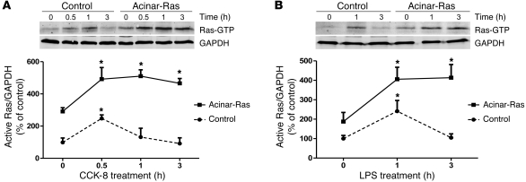
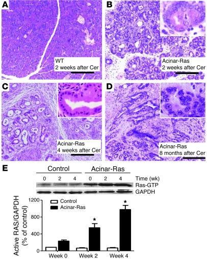
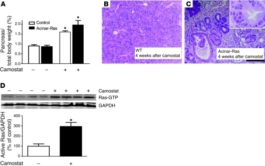
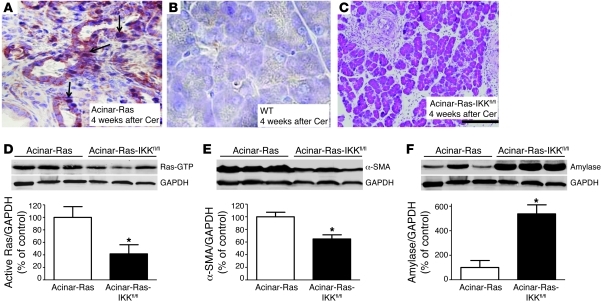
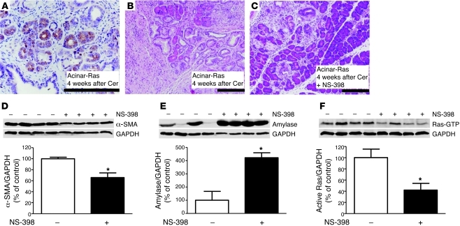
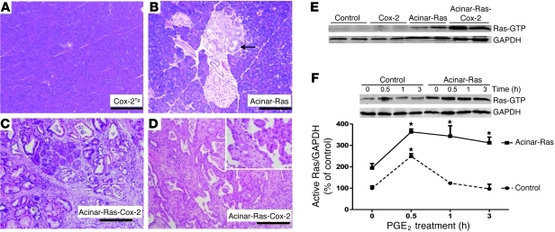
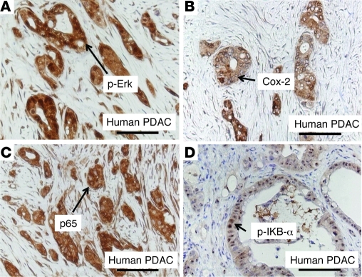
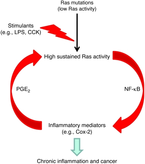
Genetic mutations that give rise to active mutant forms of Ras are oncogenic and found in several types of tumor. However, such mutations are not clear biomarkers for disease, since they are frequently detected in healthy individuals. Instead, it has become clear that elevated levels of Ras activity are critical for Ras-induced tumorigenesis. However, the mechanisms underlying the production of pathological levels of Ras activity are unclear. Here, we show that in the presence of oncogenic Ras, inflammatory stimuli initiate a positive feedback loop involving NF-κB that further amplifies Ras activity to pathological levels. Stimulation of Ras signaling by typical inflammatory stimuli was transient and had no long-term sequelae in wild-type mice. In contrast, these stimuli generated prolonged Ras signaling and led to chronic inflammation and precancerous pancreatic lesions (PanINs) in mice expressing physiological levels of oncogenic K-Ras. These effects of inflammatory stimuli were disrupted by deletion of inhibitor of NF-κB kinase 2 (IKK2) or inhibition of Cox-2. Likewise, expression of active IKK2 or Cox-2 or treatment with LPS generated chronic inflammation and PanINs only in mice expressing oncogenic K-Ras. The data support the hypothesis that in the presence of oncogenic Ras, inflammatory stimuli trigger an NF-κB-mediated positive feedback mechanism involving Cox-2 that amplifies Ras activity to pathological levels. Because a large proportion of the adult human population possesses Ras mutations in tissues including colon, pancreas, and lung, disruption of this positive feedback loop may be an important strategy for cancer prevention.


Figures

Figure 1
Figure 1. Stimuli induced prolonged increases in Ras activity in cells prepared from acinar-Ras but not control mice.
Freshly isolated acini from control or acinar-Ras mice were stimulated with 10 nM CCK-8 (A) or 1 μg/ml LPS (B) for different time periods. The GTP-bound active Ras was measured by a Raf pull-down assay. In each case, a representative Western blot is shown with quantitative data from n = 3–4 independent experiments (*P < 0.05 versus time 0).
Figure 2
Figure 2. Cerulein administration led to the development of chronic pancreatitis and PanINs and prolonged elevated Ras activity only in the presence of mutant K-Ras.
The CCK analog cerulein was injected using a scheme known to induce acute inflammation in the pancreas (Supplemental Figure 1). Pancreata from control littermates recovered from the acute effects of these injections and were histologically normal by day 14 (A, original magnification, ×100). In contrast, acinar-Ras animals showed a depletion of acinar cells, sustained edema, and inflammation 2 weeks after the first series of cerulein (Cer) treatments (B, original magnification, ×100; inset, ×400). By day 28, the pancreata from acinar-Ras mice showed abundant fibrosis and PanINs (C, original magnification, ×100; inset, ×400). Pancreatic ductal adenocarcinoma developed in acinar-Ras mice 8 months after cerulein treatments (D, original magnification, ×100; inset, ×400). For Ras activity assays, animals were sacrificed without treatment (week 0), 2 weeks after one series of cerulein injections (week 2), and 2 weeks after a second series of cerulein injections (week 4). Cerulein treatments caused a transient increase in signaling in littermate controls but a sustained and increasing level of Ras activity in acinar-Ras mice (E) (*P < 0.05 compared with controls; n = 8 animals).
Figure 3
Figure 3. Camostat feeding caused chronic pancreatitis and PanINs in acinar-Ras but not control mice.
Acinar-Ras or control mice were fed a diet containing 0.1% camostat for 4 weeks to raise endogenous levels of CCK. Pancreas growth was measured as a fraction of body weight (A) (*P < 0.05; n = 4 animals). Camostat feeding of control mice did not lead to noticeable histologic changes in the pancreas (B, original magnification, ×100). In contrast, camostat feeding of acinar-Ras mice led to the development of chronic pancreatitis and PanINs (C, original magnification, ×100; inset, ×400). Camostat feeding also caused a sustained elevation of Ras activity in the pancreata of acinar-Ras mice (D) (*P < 0.05; n = 4 animals).
Figure 4
Figure 4. NF-κB activation was essential for cerulein to induce chronic inflammation and precancerous lesions in acinar-Ras mice.
After 2 series of cerulein treatments (week 4), pancreata of acinar-Ras mice showed increased NF-κB subunit p65 nucleus translocation (A, arrows; original magnification, ×400) as compared with controls (B, ×400). Triple transgenic mice (acinar-Ras-IKKfl/fl), which express mutant K-RasG12D with IKK2 deletion, displayed a significant reduction of fibrosis and inflammation (C, original magnification, ×100) and Ras activity (D) after 2 series of cerulein treatments. The degree of stellate cell activation and retention of pancreas parenchyma in acinar-Ras-IKKfl/fl mice was measured by quantification of α-SMA for stellate cells (E) and amylase for acinar cells (F) (*P < 0.05 compared with acinar-Ras mice; n = 4 animals). GAPDH was probed for relative protein loading control (note: GAPDH in D and F was from the same gel).
Figure 5
Figure 5. NF-κB activation accelerated oncogenic Ras–induced pathologies.
Pancreata of acinar-Ras mice 2 weeks after induction of mutant K-RasG12D expression were histologically normal (A, original magnification, ×200). Pancreata of IKKTg mice showed limited inflammation (B, original magnification, ×200). In contrast, expression of both K-RasG12D and IKK2 led to dramatic fibrosis of the pancreas and the development of multiple PanINs (C, original magnification, ×200). The downstream Ras effector p-Erk was dramatically increased in the pancreas of acinar-Ras-IKKTg mice (D, original magnification, ×200) in comparison to mice expressing acinar-Ras (E, ×200) or IKK (F, ×200) alone.
Figure 6
Figure 6. Cox-2 inhibition reduced the development of cerulein-induced chronic inflammation in acinar-Ras mice.
Cerulein treatment increased Cox-2 expression in the pancreas of acinar-Ras mice (week 4) (A, original magnification, ×200) and generated chronic inflammation and fibrosis (B, ×100). Simultaneous treatment with a selective Cox-2 inhibitor (NS-398, 5 mg/kg/d) dramatically reduced the severity of these effects (week 4) (C, original magnification, ×100). These data were confirmed by quantification of decreased α-SMA (D) and increased amylase expression (E) in acinar-Ras mice with NS-398 administration. Treatment with the selective Cox-2 inhibitor also significantly decreased Ras activity (F) (*P < 0.05 versus nontreated acinar-Ras mice; n = 4 animals).
Figure 7
Figure 7. Cox-2 and oncogenic K-Ras synergized to promote the development of chronic inflammation and cancer.
Pancreata from mice expressing acinar cell Cox-2 were histologically normal at 2 months of age (A, original magnification, ×100). Similarly, pancreata from mice with expression of mutant K-Ras in acinar cells were mostly normal, although occasional low-grade PanIN lesions were observed at 2 months of age (B, arrow; original magnification, ×100). In contrast, mice expressing both Cox-2 and mutant K-Ras developed severe chronic pancreatitis at 2 months of age, with dramatic fibrosis, inflammation, destruction of acinar cells, and multiple PanINs (C, original magnification, ×100). Cancer in situ was observed (1 of 6) in K-Ras-Cox-2 mice within 6 months (D, original magnification, ×100; inset, ×400). Ras activity was greatly increased in the pancreata expressing both Cox-2 and oncogenic Ras (E). Stimulation of freshly isolated acini with 10 μM PGE2 caused a prolonged increase in Ras activity in acinar-Ras mice but not control mice (F) (*P < 0.05 versus time 0).
Figure 8
Figure 8. Ras, Cox-2, and NF-κB pathways were upregulated in human pancreatic cancer.
Increased p-Erk, an indicator of elevated Ras signaling, was observed in human cancer tissues (A). Similarly, Cox-2 expression was highly upregulated in human pancreatic cancer cells (B). p65 nuclear translocation in cancer cells induced an increase in NF-κB signaling (C). Positive staining of p–IκB-α, a direct target of IKK kinase, suggested the presence of high IKK kinase activity (D). Negative staining of these signaling molecules in normal human pancreas is shown in Supplemental Figure 8. PDAC, pancreatic ductal adenocarcinoma.
Figure 9
Figure 9. An NF-κB pathway–mediated positive feedback loop amplifies Ras activity to pathological levels in cells expressing oncogenic Ras.
Physiological levels of oncogenic Ras generate increased Ras activity but rarely lead to pathologies. However, in the presence of oncogenic Ras, inflammatory stimuli induce high sustained levels of Ras activity, which in turn induce more inflammatory mediators. This positive feedback loop amplifies and prolongs Ras activity to pathological levels that cause chronic inflammation and cancer.

Similar articles

Cited by

References

    1. DeNicola GM, Tuveson DA. RAS in cellular transformation and senescence. Eur J Cancer. 2009;45(suppl 1):211–216. doi: 10.1016/S0959-8049(09)70036-X. - DOI - PubMed
    1. Yan L, et al. Molecular analysis to detect pancreatic ductal adenocarcinoma in high-risk groups. Gastroenterology. 2005;128(7):2124–2130. doi: 10.1053/j.gastro.2005.03.006. - DOI - PubMed
    1. Lu X, Xu T, Qian J, Wen X, Wu D. Detecting K-ras and p53 gene mutation from stool and pancreatic juice for diagnosis of early pancreatic cancer. Chin Med J (Engl). 2002;115(11):1632–1636. - PubMed
    1. Parsons BL, Meng F. K-RAS mutation in the screening, prognosis and treatment of cancer. Biomark Med. 2009;3(6):757–769. doi: 10.2217/bmm.09.95. - DOI - PubMed
    1. Yakubovskaya MS, et al. High frequency of K-ras mutations in normal appearing lung tissues and sputum of patients with lung cancer. Int J Cancer. 1995;63(6):810–814. doi: 10.1002/ijc.2910630611. - DOI - PubMed

Publication types

MeSH terms