. 2012 May 1;72(9):2339-49.
doi: 10.1158/0008-5472.CAN-11-4149. Epub 2012 Mar 9.

Novel MT1-MMP small-molecule inhibitors based on insights into hemopexin domain function in tumor growth

Affiliations

Novel MT1-MMP small-molecule inhibitors based on insights into hemopexin domain function in tumor growth

Albert G Remacle et al. Cancer Res. .

Abstract

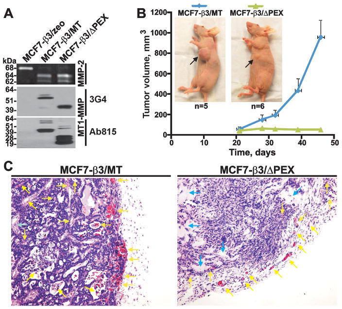
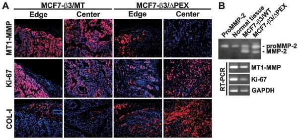
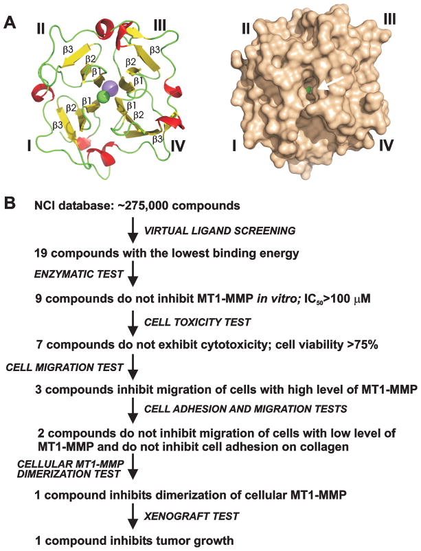
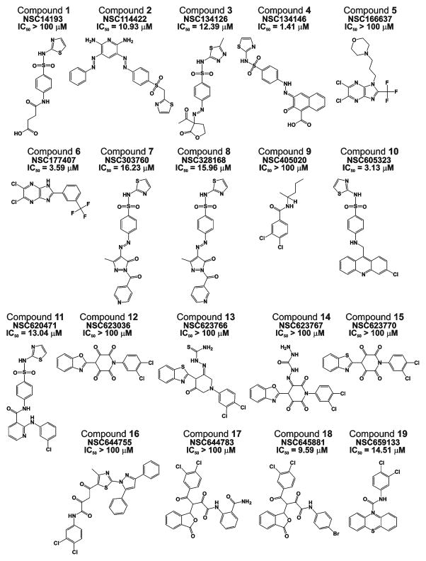
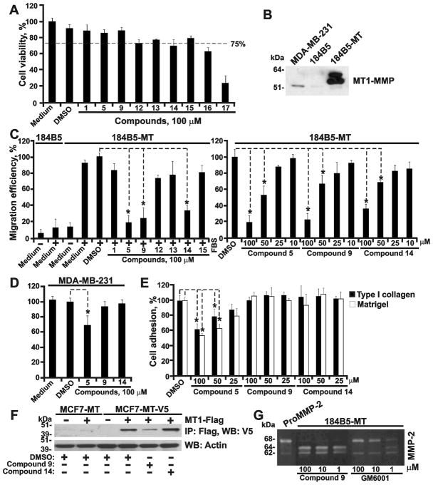
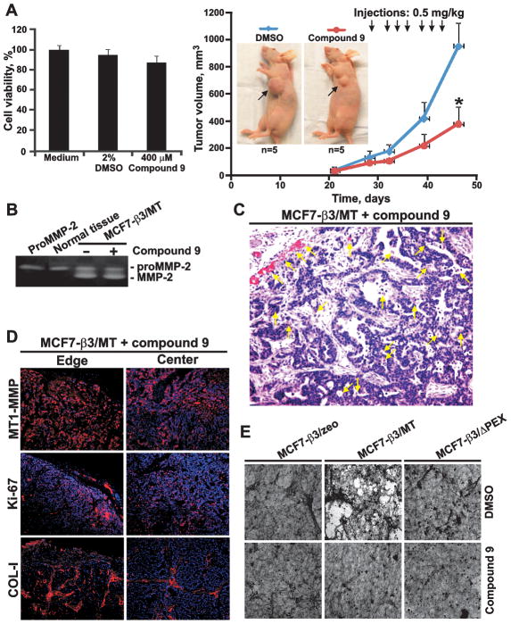
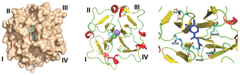
Membrane type-1 matrix metalloproteinase (MT1-MMP) is a promising drug target in malignancy. The structure of MT1-MMP includes the hemopexin domain (PEX) that is distinct from and additional to the catalytic domain. Current MMP inhibitors target the conserved active site in the catalytic domain and, as a result, repress the proteolytic activity of multiple MMPs instead of MT1-MMP alone. In our search for noncatalytic inhibitors of MT1-MMP, we compared the protumorigenic activity of wild-type MT1-MMP with an MT1-MMP mutant lacking PEX (ΔPEX). In contrast to MT1-MMP, ΔPEX did not support tumor growth in vivo, and its expression resulted in small fibrotic tumors that contained increased levels of collagen. Because these findings suggested an important role for PEX in tumor growth, we carried out an inhibitor screen to identify small molecules targeting the PEX domain of MT1-MMP. Using the Developmental Therapeutics Program (National Cancer Institute/NIH), virtual ligand screening compound library as a source and the X-ray crystal structure of PEX as a target, we identified and validated a novel PEX inhibitor. Low dosage, intratumoral injections of PEX inhibitor repressed tumor growth and caused a fibrotic, ΔPEX-like tumor phenotype in vivo. Together, our findings provide a preclinical proof of principle rationale for the development of novel and selective MT1-MMP inhibitors that specifically target the PEX domain.


Figures

Fig. 1
Fig. 1. MCF7-β3/MT and MCF7-β3/ΔPEX cells
(A) Gelatin zymography of MMP-2 (top) and Western blotting with the MT1-MMP 3G4 (middle) and Ab815 (bottom) antibodies. MCF7-β3/zeo cells, control. (B) MCF7-β3/MT and MCF7-β3/ΔPEX tumor growth in mice. Arrows indicate tumors. (C) H&E staining of tumors. Yellow and blue arrows point to blood vessels/T cells and the stroma, respectively.
Fig. 2
Fig. 2. MCF7-β3/MT and MCF7-β3/ΔPEX tumors
(A) Immunostaining of xenografts. MT1-MMP, Ki-67 and COL-I were stained with the primary antibody followed by species-specific Alexa 594-conjugated secondary antibody (red). DAPI, blue. (B) Gelatin zymograpy (top) and RT-PCR (bottom) of tumors. Left lane, top – proMMP-2 control. Normal tissue, normal mammary tissue.
Fig. 3
Fig. 3. Selection of PEX-targeting compounds
(A) Cartoon and surface representation of the four-bladed propeller structure of PEX (left and right, respectively). Blades I–IV and selected β-strands are labeled. Arrow points to the druggable pocket. Sodium atom, pink. Chlorine atom, green. (B) Screening steps and tests we used to select PEX-targeting compounds. Images were created using PyMol.
Fig. 4
Fig. 4. Top-ranking PEX-targeting compounds
NSC identifiers and IC50 values against CAT are shown.
Fig. 5
Fig. 5. Compound selection steps
(A) Cell toxicity assay using 184B5-MT cells. (B) Western blotting with the MT1-MMP 3G4 antibody. Cells were co-incubated for 24 h with GM6001. (C) Migration assay on COL-I using 184B5-MT cells. 184B5 cells, non-migratory control. Where indicated, compounds or DMSO (1%) were added to the cells. Left, 100 μM compounds. Right, 10–100 μM compounds 5, 9 and 14. (D) Migration assay on COL-I using MDA-MB-231 cells. Where indicated, compounds (100 μM) or DMSO (1%) were added to the cells. (E) Cell adhesion assay on COL-I and Matrigel using 184B5-MT cells. Compounds 5, 9 and 14 (25–100μM) or DMSO (1%) were added to cells. (F) MT1-MMP dimerization test. MCF7-MT and MCF7-MT-V5 cells transfected with the MT1-MMP-FLAG (MT1-FLAG) construct were co-incubated with compounds 9 and 14 (100 μM) or DMSO. MT1-MMP-FLAG was then immunoprecipitated (IP) using the FLAG antibody-beads and the MT1-MMP-V5 construct was measured in the FLAG-precipitates by Western blotting (WB) with the V5 antibody. Actin, loading control. (G) Gelatin zymography of 184B5-MT cells. Cells were co-incubated with purified proMMP-2, compound 9 (1–100 μM) or GM6001 control (1–100 μM). *, p<0.05.
Fig. 6
Fig. 6. Compound 9
(A) Left, Cell toxicity assay using MCF7-β3/MT. Cells were co-incubated for 6 h with compound 9 (400 μM) or DMSO (2%). Right, Effect of compound 9 on MCF7-β3/MT tumor growth. Mice received 7 injections of compound 9 (0.5 mg/kg) starting on day 29. Arrows indicate tumors. (B) Gelatin zymograpy of MCF7-β3/MT tumors. Where indicated, mice received compound 9. Left lane, proMMP-2 control. Normal tissue, normal mammary tissue. (C) H&E staining of MCF7-β3/MT tumors treated with compound 9. Yellow arrows indicate blood vessels. (D) Immunostaining of MCF7-β3/MT tumors treated with compound 9. MT1-MMP, Ki-67 and COL-I were stained with the primary antibodies followed by the Alexa 594-conjugated secondary antibody (red). DAPI, blue. (E) COL-I degradation. Cells were plated on COL-I with compound 9 or DMSO. In 3 days cells were removed. COL-I was stained using Coomassie.
Fig. 7
Fig. 7. Modeled PEX-compound 9 structure
Left, structure of a PEX-compound 9 complex. PEX is shown as molecular surface. Middle, cartoon representation of PEX with compound 9 (colored by chemical element type). Right, close-up shows residues proximal to compound 9 (blue). Sodium atom, pink. Chlorine atom, green.

References

    1. Zucker S, Pei D, Cao J, Lopez-Otin C. Membrane type-matrix metalloproteinases (MT-MMP) Curr Top Dev Biol. 2003;54:1–74. - PubMed
    1. Coussens LM, Fingleton B, Matrisian LM. Matrix metalloproteinase inhibitors and cancer: trials and tribulations. Science. 2002;295:2387–92. - PubMed
    1. Egeblad M, Werb Z. New functions for the matrix metalloproteinases in cancer progression. Nat Rev Cancer. 2002;2:161–74. - PubMed
    1. Hotary K, Allen E, Punturieri A, Yana I, Weiss SJ. Regulation of cell invasion and morphogenesis in a three-dimensional type I collagen matrix by membrane-type matrix metalloproteinases 1, 2, and 3. J Cell Biol. 2000;149:1309–23. - PMC - PubMed
    1. Hotary KB, Yana I, Sabeh F, Li XY, Holmbeck K, Birkedal-Hansen H, et al. Matrix metalloproteinases (MMPs) regulate fibrin-invasive activity via MT1-MMP-dependent and -independent processes. J Exp Med. 2002;195:295–308. - PMC - PubMed

Publication types

MeSH terms