Review
. 2012 Jul;40(12):5189-200.
doi: 10.1093/nar/gks226. Epub 2012 Mar 9.

Structural, functional and evolutionary relationships between homing endonucleases and proteins from their host organisms

Affiliations
Review

Structural, functional and evolutionary relationships between homing endonucleases and proteins from their host organisms

Gregory K Taylor et al. Nucleic Acids Res. 2012 Jul.

Abstract

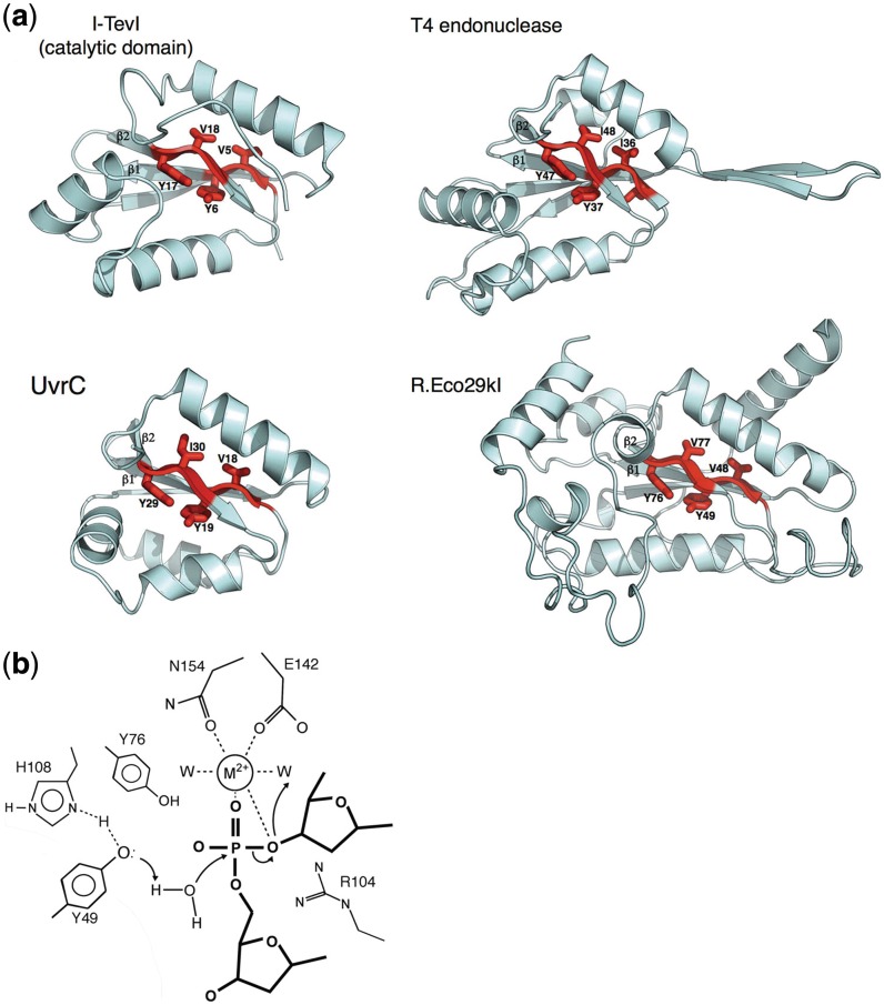
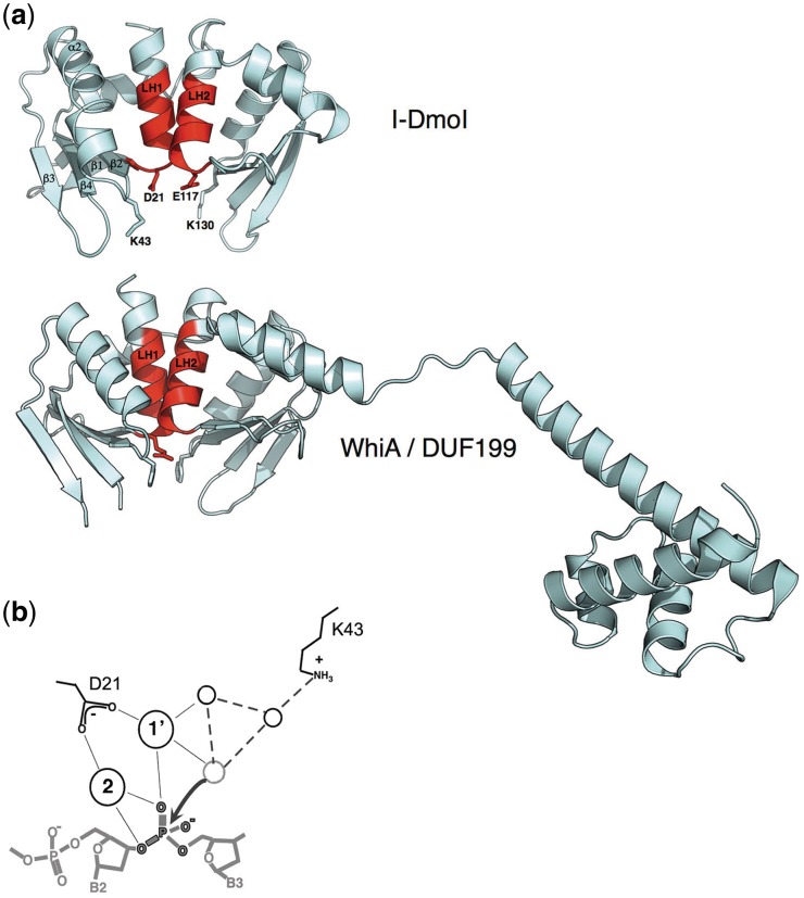
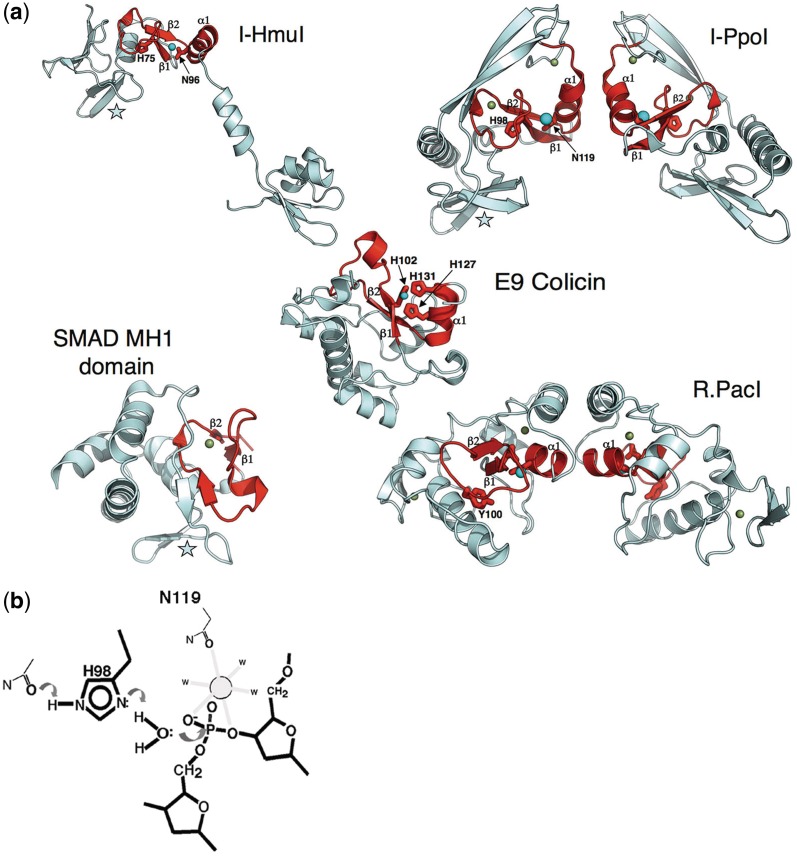
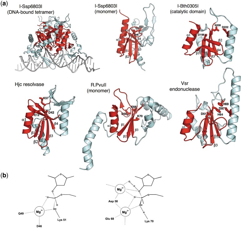
Homing endonucleases (HEs) are highly specific DNA-cleaving enzymes that are encoded by invasive DNA elements (usually mobile introns or inteins) within the genomes of phage, bacteria, archea, protista and eukaryotic organelles. Six unique structural HE families, that collectively span four distinct nuclease catalytic motifs, have been characterized to date. Members of each family display structural homology and functional relationships to a wide variety of proteins from various organisms. The biological functions of those proteins are highly disparate and include non-specific DNA-degradation enzymes, restriction endonucleases, DNA-repair enzymes, resolvases, intron splicing factors and transcription factors. These relationships suggest that modern day HEs share common ancestors with proteins involved in genome fidelity, maintenance and gene expression. This review summarizes the results of structural studies of HEs and corresponding proteins from host organisms that have illustrated the manner in which these factors are related.


Figures

Figure 1.
Figure 1.
The GIY-YIG motif. (a) Enzymes involved in homing (I-TevI), DNA degradation (T4 endonuclease), restriction (R.Eco29kI) and DNA repair (UvrC) contain similar GIY-YIG catalytic cores, in which two short antiparallel β-strands contain the conserved signature residues for the enzyme family (shown in red). Based on crystal structures of the R.Eco29kI and R.Hpy188I REases bound to their DNA targets, the two strongly conserved tyrosine residues in the catalytic motif (Y6 and Y17 in I-TevI) are believed to be involved in general acid–base catalysis and activation of a water nucleophile (13,14). (b) The putative catalytic mechanism of GIY-YIG endonucleases involves activation of incoming nucleophilic water by an active-site tyrosine, which is itself activated through interactions with surrounding basic side chains. A single-bound divalent metal ion is coordinated by the scissile phosphate and neighboring active site side chains. This mechanism and active site bears a strong resemblance to the HNH nuclease motif (Figure 3), but has evolved using a completely different surrounding protein-fold topology. The side chain labels and features shown are based on the R.Eco29kI/DNA crystal structure (13).
Figure 2.
Figure 2.
The LAGLIDADG motif. (a) HEs such as I-DmoI display a core fold consisting of two copies of an ‘αββαββ’ topology in which the first helix in each fold (colored red and labeled ‘LH1’ and ‘LH2’) contain the consensus sequence motif, and pack against one another to comprise both a domain interface and the center of the endonuclease active site. An acidic residue (usually an aspartate, but in many cases, a glutamate) extends from the bottom of each helix (D21 and E117 in I-DmoI); together they coordinate multiple divalent metal ions in conjunction with the scissile phosphate oxygens. Strongly conserved basic residues (K43 and K130 in I-DmoI) extend from the β1−β2 loop in the active sites and are believed to play a role in stabilizing the phosphoanion transition state and/or assisting in general acid/base catalysis. In contrast, the WhiA/DUF199 family of bacterial gene regulators contains a LAGLIDADG protein domain that closely resembles the HE structural family, tethered to a C-terminal helix-turn-helix domain. However, the catalytic acidic and basic residues described above are not conserved (in the case of the WhiA protein from Thermatogus maritima, the positions of the LAGLIDADG acidic residues are instead an arginine and glycine; the positions of the neighboring basic residues are two phenylalanines. As well, the overall positively charged surface of the HE that is formed by its β-sheets is instead considerably more varied in its charge composition, indicating that the DNA-binding properties of the LAGLIDADG fold have been replaced with alternative roles. (b) The putative mechanism of the LAGLIDADG endonucleases involves activation of incoming metal-bound nucleophilic water by a network of surrounding basic side chains and additional solvent molecules. The two most conserved residues in the active site (indicated and labeled based on the structure of the I-DmoI endonuclease) are an acidic metal-binding residue contributed by the penultimate residue of each LAGLIDADG motif and a neighboring basic residue (usually a lysine) bound on an adjacent DNA-binding loop. Cleavage of the DNA appears to follow a mechanism that involves two bound metals for each DNA-strand scission event. Many LAGLIDADG endonucleases display considerable disparity in the kinetics of individual strand cleavage events, such that significant fraction of nicked intermediate accumulates prior to final double-strand break formation.
Figure 3.
Figure 3.
The ββα−metal motif. (a) HE families found in either phage (such as I-HmuI) or in protists (such as I-PpoI) contain quite similar ββα-metal catalytic core folds and active-site residues (colored red). In both enzymes, an active-site histidine residue (H75 in I-HmuI; H98 in I-PpoI) acts as a general base to assist in deprotonation and activation of an active-site water molecule. A neighboring asparagine located at the N-terminal end of the motif's α-helix is involved in coordination of a single-bound divalent metal ion, which participates in transition state stabilization. Both active-site core folds are embellished by a small antiparallel β-sheet (denoted by a star), which is involved in sequence-specific DNA-site recognition, by forming base pair interactions in the target site major groove. Beyond these common elements, the two HEs differ significantly. I-HmuI displays an extended monomeric structure in which a pair of α-helices forms additional contacts to the 3′ end of the DNA-target site. In contrast, I-PpoI forms a homodimeric structure in which two identical copies of the enzyme DNA-binding surface interact with distal ends of a symmetric DNA sequence. The ββα-metal core fold is found in proteins with substantially different biochemical functions and biological roles, ranging from competitive DNA degradation and toxicity (the bacterial colicins), phage restriction (R.PacI), and even eukaryotic transcriptional regulation (the SMAD proteins). The divergent biological roles of these proteins is reflected in additional sequence and structural variation of the ββα-metal core: the colicins display considerably different metal coordination schemes from the HEs (E9 colicin employs three histidine residues); R.PacI appears to have replaced the active-site histidine base with a tyrosine, and the DNA-binding MH1 domain of the SMAD proteins have completely devolved catalytic regions, but have maintained the same overall architecture of the DNA-binding surface (also indicated with a star) as is observed for I-HmuI and I-PpoI. (b)The putative mechanisms of DNA cleavage by the ββα-metal nucleases involves activation of an incoming nucleophilic water by an active-site histidine, which is itself activated through interactions with surrounding basic side chains. A single-bound divalent metal ion is coordinated by the scissile phosphate and neighboring active site side chains. This mechanism and active site bears a strong resemblance to the HNH nuclease motif (Figure 1), but has evolved using a completely different surrounding protein-fold topology. The side chain labels and features shown are based on the I-PpoI/DNA crystal structure (10).
Figure 4.
Figure 4.
The PD-(D/E)xK motif. (a)As has been observed for the ββα-metal motif (Figure 3), HEs found in two widely different genomic niches (phage and eubacteria) have evolved that contain different versions of the PD-(D/E)xK nuclease motif. In the traditional motif (I-Ssp6803I and its closest structural relatives), a minimum of two acidic residues and one basic residue are position within a mixed α/β topology (colored in red) and participate in divalent metal coordination and promote general acid/base catalysis and/or transition state stabilization. The I-Ssp6803I HE (found within a cyanobacterium) forms a tetrameric structure in which two copies of the enzyme fold contact the DNA-target half-sites, while another two copies promote the overall quaternary interactions necessary to properly position the two binding surfaces at opposite ends of the target. Similar subunit architectures and active site chemistries are observed both for many different DNA-active enzymes. Some of the mostly closely related such enzymes to the I-Ssp6803I HE, identified using automated searches of the PDB database, are an archael Holliday junction resolvase (Hjc) and the R.PvuII and R.SfiI REases. In contrast, the phage-derived I-Bth0305I HE displays a common core-folded topology as the traditional PD-(D/E)xK nucleases, but displays a significantly diverged active-site architecture and presumed mechanism of DNA cleavage that most closely resembles the very short patch repair (Vsr) endonuclease (42). Such HEs have been termed the ‘EDxHD’ family (3) to denote their conservation pattern of active-site residues and to distinguish them from the bacterial HEs.(b) The generic mechanism of DNA hydrolysis by the PD-(D/E)xK containing nucleases involves the activation of a metal-bound water either through direct interaction with a basic side chain, which acts as a general base, or through a water-mediated contact. PD-(D/E)xK nucleases display considerable variation both in the number and exact position of bound metal ions during catalysis, as well as the exact structural position within the core protein fold of each catalytic residue. The I-Ssp6803I HE appears to bind one divalent metal ion (left), while the R.PvuII REase is thought to bind metal ions within at least two distinct sites during catalysis (right).

References

    1. Dujon B. Group I introns as mobile genetic elements: facts and mechanistic speculations – a review. Gene. 1989;82:91–114. - PubMed
    1. Stoddard BL. Homing endonucleases: from microbial genetic invaders to reagents for targeted DNA modification. Structure. 2011;19:7–15. - PMC - PubMed
    1. Taylor GK, Heiter DF, Pietrokovski S, Stoddard BL. Activity, specificity and structure of I-Bth0305I: a representative of a new homing endonuclease family. Nucleic Acids Res. 2011;39:9705–9719. - PMC - PubMed
    1. Dunin-Horkawicz S, Feder M, Bujnicki JM. Phylogenomic analysis of the GIY-YIG nuclease superfamily. BMC Genomics. 2006;7:98. - PMC - PubMed
    1. Dalgaard JZ, Klar AJ, Moser MJ, Holley WR, Chatterjee A, Mian IS. Statistical modeling and analysis of the LAGLIDADG family of site-specific endonucleases and identification of an intein that encodes a site-specific endonuclease of the HNH family. Nucleic Acids Res. 1997;25:4626–4638. - PMC - PubMed

Publication types

MeSH terms