. 2012 Jul;40(12):5402-14.
doi: 10.1093/nar/gks224. Epub 2012 Mar 9.

Drosophila melanogaster linker histone dH1 is required for transposon silencing and to preserve genome integrity

Affiliations

Drosophila melanogaster linker histone dH1 is required for transposon silencing and to preserve genome integrity

Olivera Vujatovic et al. Nucleic Acids Res. 2012 Jul.

Abstract

Histone H1 is an intrinsic component of chromatin, whose important contribution to chromatin structure is well-established in vitro. Little is known, however, about its functional roles in vivo. Here, we have addressed this question in Drosophila, a model system offering many advantages since it contains a single dH1 variant. For this purpose, RNAi was used to efficiently deplete dH1 in flies. Expression-profiling shows that dH1 depletion affects expression of a relatively small number of genes in a regional manner. Furthermore, depletion up-regulates inactive genes, preferentially those located in heterochromatin, while active euchromatic genes are down-regulated, suggesting that the contribution of dH1 to transcription regulation is mainly structural, organizing chromatin for proper gene-expression regulation. Up-regulated genes are remarkably enriched in transposons. In particular, R1/R2 retrotransposons, which specifically integrate in the rDNA locus, are strongly up-regulated. Actually, depletion increases expression of transposon-inserted rDNA copies, resulting in synthesis of aberrant rRNAs and enlarged nucleolus. Concomitantly, dH1-depleted cells accumulate extra-chromosomal rDNA, show increased γH2Av content, stop proliferation and activate apoptosis, indicating that depletion causes genome instability and affects proliferation. Finally, the contributions to maintenance of genome integrity and cell proliferation appear conserved in human hH1s, as their expression rescues proliferation of dH1-depleted cells.


Figures

Figure 1.
Figure 1.
dH1 depletion has a dual effect on gene expression. (A) The number of genes differentially expressed in mutant his1RNAi; Act5C-GAL4 flies is presented as a function of increasing fold-change (FC) for both up- (red) and down-regulated genes (blue). (B) The overlapping between genes differentially expressed (|FC| > 1.5) in mutant his1RNAi; Act5C-GAL4 flies (red), and dH1-rich regions in S2 (blue) and/or BG3 cells (black) is presented. (C) The average expression in control GFPRNAi; Act5C-GAL4 flies of genes differentially up- (red) and down-regulated (blue) in mutant his1RNAi; Act5C-GAL4 flies is presented for the indicated FCs. (D) The average fold change expression in mutant his1RNAi; Act5C-GAL4 flies is presented for the ±40 genes immediately flanking genes highly differentially expressed (FC > 2.5). Up- (red) and down-regulated (blue) genes are indicated. The ratios of the number of up-regulated versus down-regulated genes in the indicated regions are presented.
Figure 2.
Figure 2.
dH1 is required to maintain silencing of heterochromatic genes and transposons. (A) The percentages of heterochromatic and euchromatic genes analyzed in the array that are found differentially expressed in mutant his1RNAi; Act5C-GAL4 flies are presented for the indicated FCs. Up- (red) and down-regulated genes (blue) are indicated. (B) Gene Set Enrichment Analysis (GSEA) for heterochromatic genes. The distribution of heterochromatic genes (black lines) is presented as a function of the change in expression in mutant his1RNAi; Act5C-GAL4 knockdown flies, from highly up-regulated (red) to highly down-regulated (blue). Enrichment score is calculated as described under ‘Materials and Methods’ section. (C) As in B but for transposons.
Figure 3.
Figure 3.
dH1 depletion deregulates expression of the rDNA locus. (A) Quantitative RT-PCR analysis of the levels of expression of R1 retrotransposon, and R1- and R2-inserted 28rRNA copies in mutant his1RNAi; Act5C-GAL4 and control GFPRNAi; Act5C-GAL4 larvae. Total RNA was prepared from wing imaginal discs. Relative expression levels were determined in relation to Rp49 mRNA. Fold change of expression in his1RNAi versus control is presented. See Supplementary Table S3 for primers used in these experiments. (B) Nucleolar morphology was determined by immunostaining with αfibrillarin (1:1000; green) in whole salivary glands prepared from mutant his1RNAi; Act5C-GAL4 and control GFPRNAi; Act5C-GAL4 larvae. DNA was stained with DAPI. (C) As in B, but immunostaining was performed in wing imaginal discs prepared from mutant his1RNAi; nub-GAL4; UASGAL4-Dcr2 and control GFPRNAi; nub-GAL4; UASGAL4-Dcr2 larvae. The region corresponding to the pouch is indicated. DNA was stained with DAPI. Enlarges images of the indicated regions are shown on the right for easier visualization. (D) Fibrillarin content was analyzed by Western blot using αfibrillarin antibodies (1:1000) in extracts prepared from mutant his1RNAi; Act5C-GAL4 and control GFPRNAi; Act5C-GAL4 salivary glands. Two increasing amounts of extract were analyzed in each case (lanes 1, 2). The signal obtained with αActin antibodies (1:750) was used as loading control for normalization.
Figure 4.
Figure 4.
dH1 depletion causes genomic instability. (A) Quantitative PCR analysis of the levels of eccDNA originated from the rDNA, Rp49 or stellate loci in mutant his1RNAi; Act5C-GAL4 and control GFPRNAi; Act5C-GAL4 larvae. Hirt extracts were prepared from imaginal discs (top) and salivary glands (bottom). Fold increase in mutant his1RNAi versus control is presented. Similar analyses performed using homozygous su(var)3-906 larvae are presented as positive controls for comparison. See Supplementary Table S3 for primers used in these experiments. (B) γH2Av levels were determined by immunostaining using αγH2Av antibodies (1:1000; red) in wing imaginal discs prepared from mutant his1RNAi; nub-GAL4; UASGAL4-Dcr2 and control GFPRNAi; nub-GAL4; UASGAL4-Dcr2 larvae. The region corresponding to the pouch is indicated. DNA was stained with DAPI.
Figure 5.
Figure 5.
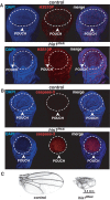
dH1-depleted cells stop proliferation and activate apoptosis. (A) The frequency of mitotic cells was determined by immunostaining using αH3S10P antibodies (1:1000; red) in wing imaginal discs prepared from mutant his1RNAi; nub-GAL4; UASGAL4-Dcr2 and control GFPRNAi; nub-GAL4; UASGAL4-Dcr2 larvae. The region corresponding to the pouch is indicated. DNA was stained with DAPI. (B) Induction of apoptosis was determined by immunostaining using αcaspase-3 antibodies (1:1000; red) in wing imaginal discs prepared from mutant his1RNAi; nub-GAL4; UASGAL4-Dcr2 and control GFPRNAi; nub-GAL4; UASGAL4-Dcr2 larvae. The region corresponding to the pouch is indicated. DNA was stained with DAPI. (C) Wings from adult mutant his1RNAi; nub-GAL4; UASGAL4-Dcr2 (right) and control GFPRNAi; nub-GAL4; UASGAL4-Dcr2 flies (left) are presented. In these experiments, his1RNAi flies carry a single UASGAL4-hsRNAHis1 construct inserted in the X-chromosome.
Figure 6.
Figure 6.
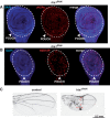
Blocking apoptosis by p35 over-expression rescues proliferation of dH1-depleted cells. (A) γH2Av levels were determined by immunostaining using αγH2Av antibodies (1:1000; red) in wing imaginal discs prepared from mutant his1RNAi; nub-GAL4; UASGAL4-Dcr2; UASGAL4-p35. The region corresponding to the pouch is indicated. DNA was stained with DAPI. (B) The frequency of mitotic cells was determined by immunostaining using αH3S10P antibodies (1:1000; red) in wing imaginal discs prepared from mutant his1RNAi; nub-GAL4; UASGAL4-Dcr2; UASGAL4-p35 larvae. The region corresponding to the pouch is indicated. DNA was stained with DAPI. (C) Wings from adult mutant his1RNAi; nub-GAL4; UASGAL4-Dcr2; UASGAL4-p35 (right) and control GFPRNAi; nub-GAL4; UASGAL4-Dcr2; UASGAL4-p35 flies (left) are presented. Red arrows indicate tumour-like outgrowths. In these experiments, his1RNAi flies carry a single UASGAL4-hsRNAHis1 construct inserted in the X-chromosome.
Figure 7.
Figure 7.
Expression of human hH1 variants rescues proliferation of dH1-depleted cells. (A) Levels of expression of HA-tagged hH1.0, hH1.2 and hH1.4 were determined by Western blot using αHA antibodies (1:500) in wing imaginal discs extracts prepared from mutant his1RNAi; nub-GAL4; UASGAL4-Dcr2 larvae carrying UASGAL4-constructs to over-express the indicated hH1 variant. Two increasing amounts of extract were analyzed in each case (lanes 1, 2). The signal obtained with αTubulin antibodies (1:2000) was used as loading control for normalization. The extent of dH1 depletion (%) determined by Western blot analysis is indicated. (B) Wings from adult mutant his1RNAi; nub-GAL4; UASGAL4-Dcr2 flies carrying UASGAL4-constructs to express the indicated hH1 variant or none (no hH1) are presented. (C) Average wing length is presented for mutant his1RNAi; nub-GAL4; UASGAL4-Dcr2 flies carrying UASGAL4-constructs to express the indicated hH1 variant or none (no hH1), and for control GFPRNAi; nub-GAL4; UASGAL4-Dcr2 flies. (D) Percentage of flies showing wings of the indicated lengths is presented for mutant his1RNAi; nub-GAL4; UASGAL4-Dcr2 flies carrying UASGAL4-constructs to express the indicated hH1 variant or not (no hH1), and for control GFPRNAi; nub-GAL4; UASGAL4-Dcr2 flies. In these experiments, his1RNAi flies carry a single UASGAL4-hsRNAHis1 construct inserted in the X-chromosome.

References

    1. Bassett A, Cooper S, Wu C, Travers A. The folding and unfolding of eukaryotic chromatin. Curr. Opin. Genet. Dev. 2009;19:159–165. - PubMed
    1. Kasinky HE, Lewis JD, Dacks JB, Ausio J. Origin of H1 linker histones. FASEB J. 2001;15:34–42. - PubMed
    1. Ramakrishnan V. Histone H1 and chromatin higher-order structure. Crit. Rev. Eukaryot. Gene Expr. 1997;7:215–230. - PubMed
    1. Ramakrishnan V. Histone structure and the organization of the nucleosome. Annu. Rev. Biophys. Biomol. Struct. 1997;26:83–112. - PubMed
    1. Robinson PJJ, Rhodes D. Structure of the ‘30 nm’ chromatin fibre: a key role for the linker histone. Curr. Opin. Struct. Biol. 2006;16:336–343. - PubMed

Publication types