. 2012 Mar 11;9(4):391-5.
doi: 10.1038/nmeth.1929.

Controlling gene expression with the Q repressible binary expression system in Caenorhabditis elegans

Affiliations

Controlling gene expression with the Q repressible binary expression system in Caenorhabditis elegans

Xing Wei et al. Nat Methods. .

Abstract

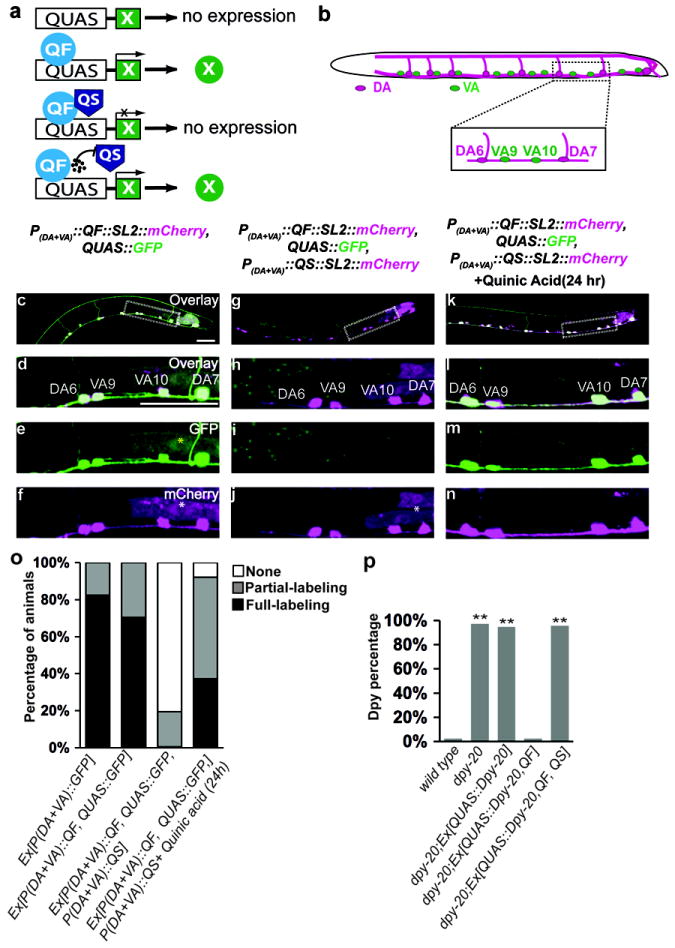
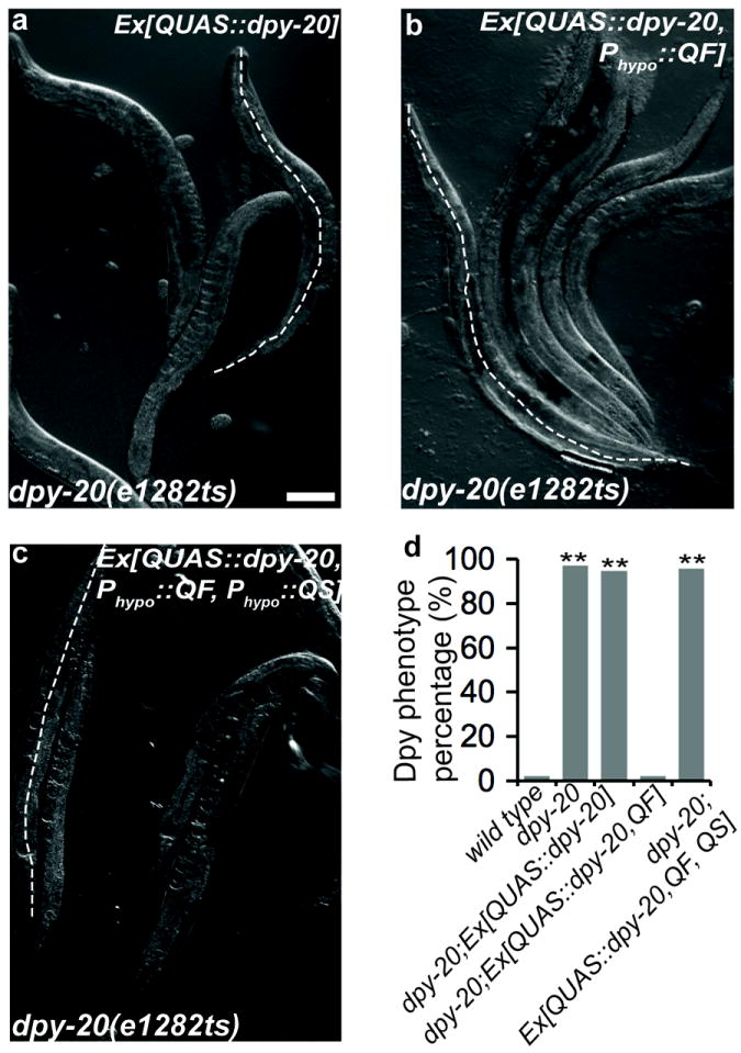
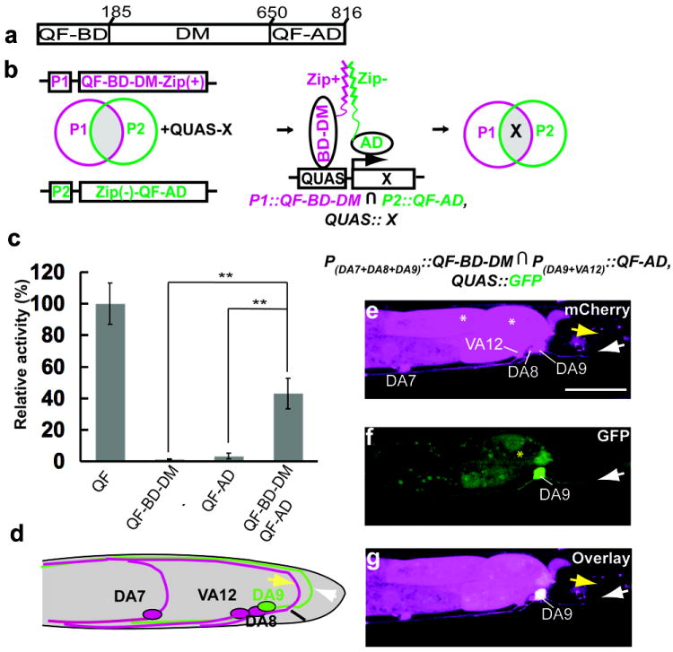
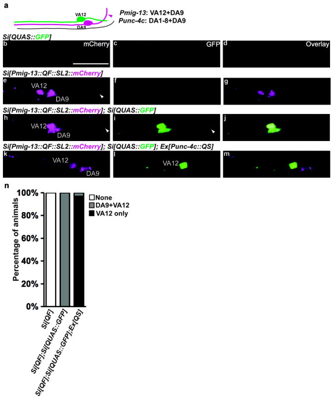
We established a transcription-based binary gene expression system in Caenorhabditis elegans using the recently developed Q system. This system, derived from genes in Neurospora crassa, uses the transcriptional activator QF to induce the expression of target genes. Activation can be efficiently suppressed by the transcriptional repressor QS, and suppression can be relieved by the nontoxic small molecule quinic acid. We used QF, QS and quinic acid to achieve temporal and spatial control of transgene expression in various tissues in C. elegans. We also developed a split Q system, in which we separated QF into two parts encoding its DNA-binding and transcription-activation domains. Each domain showed negligible transcriptional activity when expressed alone, but expression of both reconstituted QF activity, providing additional combinatorial power to control gene expression.


Conflict of interest statement

Competing Financial Interests

The authors declare no competing financial interests.

Figures

Fig. 1
Fig. 1. The repressible Q binary system functions effectively in C. elegans
(a) Schematic of the Q system. X indicates transgene. Black circles at the bottom indicate quinic acid. (b) Schematic diagram of VA and DA motor neurons in the ventral nerve cord (VNC). The boxed region is magnified to show VA9 and VA10 neurons in green and DA6 and DA7 in magenta. (c-n) The micrographs show transgenic worms (L3 larvae) expressing the indicated transgenes, with or without treatment with quinic acid for 24h. An overview of the VNC (c, g, k) and magnification of the boxed region (d-n) are shown. GFP expression is shown green, mCherry expression is shown in magenta. P, promoter; SL2, trans-spliced leader sequence. White asterisks denote ectopic gut fluorescence caused by SL2∷mCherry cassette (X. Wei and K. Shen, unpublished results). Yellow asterisks denote occasional ectopic gut fluorescence due to QUAS∷GFP. Scale bars, 20 μm. (o) Quantification of labeling efficiency in (c-n). Late L3 or early L4 stage larvae were scored (n>200 for each strain). Animals are divided into 3 categories: None (no A-type neurons labeled), Full-labeling (all A-type neurons labeled) and Partial-labeling (between None and Full-labeling). (p) Quantification of Dpy rescue efficiency with Q system in Supplementary Figure 4. n =40 for each group; **P ≤0.0001 (versus wild type animals), χ2 test. Animals with a body length longer than 972 μm are scored as wild type. Animals with a body length shorter than 972 μm are scored as Dpy mutants.
Fig. 2
Fig. 2. Refining expression patterns in VA motor neurons with a NOT gate
(a) Schematic diagram of VA and DA motor neurons in the posterior region of the VNC. DA neurons extend axonal commissures (magenta arrowheads) to the dorsal nerve cord. The unc-4 promoter is expressed in VA and DA neurons while the unc-4c promoter is only expressed in DA neurons. (b-d) The micrographs show the same region as schematically depicted in (a) in an early L4 larva expressing the indicated transgenes. Magenta shows mCherry expression and green shows GFP expression. White arrowheads indicate the commissures of DA neurons. The asterisk denotes ectopic gut fluorescence caused by SL2∷mCherry cassette. Scale bar, 20 μm.
Fig. 3
Fig. 3. The Split Q system
(a) Schematic showing the DNA binding domain (QF-BD), the putative dimerization domain (DM) and the transcription activation domain (QF-AD) of QF. The amino acid positions for each domain are indicated. (b) Schematic depiction of the Split Q system. The QF-BD-DM and QF-AD are driven using different promoters (P1 and P2). Zip+ and Zip- are a pair of heterodimerization leucine zippers. The transgene X is expressed only at the intersection of P1 and P2 promoters (gray area). ∩ denotes the intersection of P1 and P2. (c) Relative transcriptional activities (measured as GFP fluorescence intensity) of the indicated Split Q constructs, normalized to the activity measured in strains containing intact QF. All constructs were driven by the same promoter (mig-13). Error bars, S.E.M. **P<0.01, n=40, one-tailed t-test. (d) Schematic diagram of DA and VA neurons in the tail region (left view). The mig-13 promoter is expressed in DA9 and VA12 neurons while the unc-4c promoter is expressed in DA7, DA8 and DA9 neurons. The yellow and white arrows indicate the commissures of DA8 and DA9 respectively. (e-g) The micrographs show an early stage 4 larva expressing the indicated transgenes. Green shows GFP fluorescence, magenta shows mCherry fluorescence. The yellow and white arrows indicate the commissures of DA8 and DA9 respectively. White asterisks denote ectopic gut fluorescence caused by SL2∷mCherry. The yellow asterisk denotes occasional ectopic gut fluorescence due to QUAS∷GFP. Scale bar, 20 μm.
Fig. 4
Fig. 4. The Q system functions effectively with single copy transgene
(a) Schematic diagram of DA9 and VA12 neurons in the tail region. The mig-13 promoter expresses in DA9 (in magenta) and VA12 (in green) neurons while the unc-4c promoter expresses in the DA9 neuron. The magenta arrowhead indicates the commissure of DA9. (b-m) The images show the same region as schematically depicted in (a) in transgenic worms (L4 larvae) containing the indicated transgenes. GFP expression is shown green and mCherry expression is shown in magenta. Si, single insertion. The white arrowheads indicate the commissures of DA9. Scale bar, 20 μm. (n) Quantification of labeling efficiency in (b-m). Late L3 or early L4 stage larvae were scored (n>100 for each strain). Animals are divided into 3 categories: None (neither DA9 nor VA12 neurons are labeled by GFP), DA9+VA12 (both neurons are labeled), VA12 only (only VA12 neuron is labeled).

References

    1. Gossen M, Bujard H. Tight Control of Gene-Expression in Mammalian-Cells by Tetracycline-Responsive Promoters. P Natl Acad Sci USA. 1992;89:5547–5551. - PMC - PubMed
    1. Brand AH, Perrimon N. Targeted Gene-Expression as a Means of Altering Cell Fates and Generating Dominant Phenotypes. Development. 1993;118:401–415. - PubMed
    1. Lee T, Luo LQ. Mosaic analysis with a repressible cell marker for studies of gene function in neuronal morphogenesis. Neuron. 1999;22:451–461. - PubMed
    1. Macosko EZ, et al. A hub-and-spoke circuit drives pheromone attraction and social behaviour in C. elegans. Nature. 2009;458:1171–1175. - PMC - PubMed
    1. Davis MW, Morton JJ, Carroll D, Jorgensen EM. Gene activation using FLP recombinase in C. elegans. Plos Genet. 2008;4:e1000028. - PMC - PubMed

Publication types

MeSH terms