. 2012 Oct;61(10):1721-33.
doi: 10.1007/s00262-012-1237-1. Epub 2012 Mar 11.

Targeting of 4-1BB by monoclonal antibody PF-05082566 enhances T-cell function and promotes anti-tumor activity

Affiliations

Targeting of 4-1BB by monoclonal antibody PF-05082566 enhances T-cell function and promotes anti-tumor activity

Timothy S Fisher et al. Cancer Immunol Immunother. 2012 Oct.

Abstract

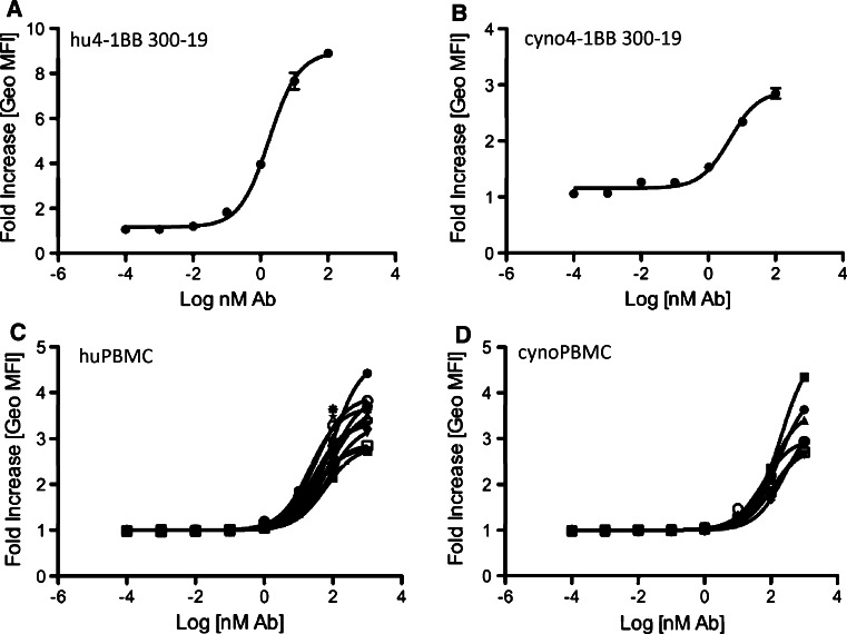
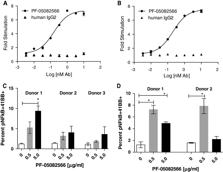
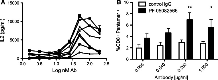
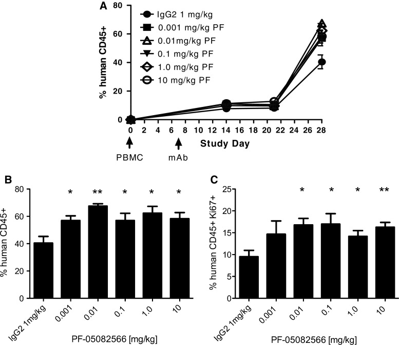
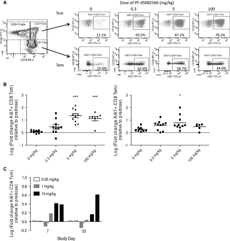
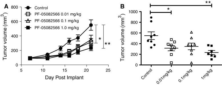
4-1BB (CD137, TNFRSF9) is a costimulatory receptor expressed on several subsets of activated immune cells. Numerous studies of mouse and human T cells indicate that 4-1BB promotes cellular proliferation, survival, and cytokine production. 4-1BB agonist mAbs have demonstrated efficacy in prophylactic and therapeutic settings in both monotherapy and combination therapy tumor models and have established durable anti-tumor protective T-cell memory responses. PF-05082566 is a fully human IgG2 that binds to the extracellular domain of human 4-1BB with high affinity and specificity. In preclinical studies, this agonist antibody demonstrated its ability to activate NF-κB and induce downstream cytokine production, promote leukocyte proliferation, and inhibit tumor growth in a human PBMC xenograft tumor model. The mechanism of action and robust anti-tumor efficacy of PF-05082566 support its clinical development for the treatment of a broad spectrum of human malignancies.


Figures

Fig. 1
Fig. 1
Saturation binding of PF-05082566 to human and cynomolgus monkey 4-1BB. Dose-dependent binding of AlexaFluor647-labeled PF-05082566 to a 300-19 cells transduced to express human 4-1BB, b 300-19 cells transduced to express cynomolgus monkey 4-1BB, c 72-h PHA-stimulated primary human PBMC, and d 72-h PHA-stimulated primary cynomolgus monkey PBMC. The data are expressed as fold increase in geometric mean fluorescence intensity (Geo MFI) of cells stained with PF-05082566 relative to cells stained with the corresponding concentration of human IgG2 isotype control as measured by FACS. a, b display data from representative experiments with samples stained in duplicate. Error bars represent SEM. c, d represent data of binding to 72-h PHA-stimulated CD3-positive primary PBMC. Each line represents the staining result of cells isolated from individual donors. Representative examples of flow cytometry data are demonstrated in Supplementary Figure 1 for 300-19 cells and Supplementary Figure 2 for PBMC
Fig. 2
Fig. 2
PF-05082566 induces NF-κB pathway activation through both human and cynomolgus monkey 4-1BB. Dose-dependent activation of an NF-κB luciferase reporter in a 293T-cells-expressing human 4-1BB or b 293T-cells-expressing cynomolgus monkey 4-1BB. The data are representative of three separate experiments and shows fold increase in luciferase activity over unstimulated control cells by goat anti-huIgG Fc-specific F(ab′)2 cross-linking antibody plus PF-05082566 (black circles) or cross-linked human IgG2 isotype control (black triangles). Additionally, PF-05082566 treatment induces phospho-NF-κB p65 in anti-CD3-stimulated primary c human and d cynomolgus monkey CD3+ cells. The graphs demonstrate percentage of phospho-NF-κB p65+, 4-1BB+ T cells as measured by FACS. Cells were stimulated with CD3 alone (white bars), CD3 plus cross-linked 0.5 μg/ml PF-05082566 (gray bars), or CD3 plus cross-linked 5 μg/ml PF-05082566 (black bars). Significance relative to the control group was determined via two-tailed Student’s t test (*p < 0.05)
Fig. 3
Fig. 3
PF-05082566 enhances IL-2 production by primary human T cells and expansion of antigen-specific CD8+ T cells in vitro. a Dose-dependent enhancement of IL-2 production as measured by ELISA from purified T cells stimulated with plate bound CD3 plus increasing concentrations of plate bound PF-05082566. Each line represents stimulation of purified T cells from one of seven individual donors tested in duplicate. b PBMC isolated from four separate HLA-A2+ donors whom had received an influenza vaccine within the prior season were stimulated in vitro with influenza matrix peptide (58–66, GILGFVFTL) for 8 days in the presence of either huIgG2 control antibody (white bars) or PF-05082566 (black bars). The data are represented as the percent viable CD8+, MHC HLA-A2-GILGFVFTL pentamer+ cells as measured by FACS. Significance was determined via 2-way ANOVA comparison with the control IgG2-treated group (**p < 0.01, *p < 0.05). Results are representative of two separate experiments using four different donors
Fig. 4
Fig. 4
PF-05082566 induces expansion of human CD45+ PBMC in vivo. Engraftment of human PBMC adoptively transferred via intraperitoneal injection to NSG host mice was analyzed by FACS for human CD45 and Ki-67 staining 28 days following injection. On day 7 following PBMC injection, mice were treated with a single injection of human IgG2 isotype control or the indicated concentration of PF-05082566. a Mean percentage of human CD45+ lymphocytes assessed at several time points over the course of the study b percentage of human CD45+ lymphocytes at Study Day 28, 21 days following mAb injection and c percentage of human CD45+ Ki-67+ present in the whole blood samples of each treatment group at Study Day 28, 21 days following mAb injection. Data are representative of n = 7 mice per group and significance relative to the control group on the final day of the study was determined via two-tailed Student’s t test (*p < 0.05, **p < 0.005)
Fig. 5
Fig. 5
Intravenous administration of PF-05082566 to cynomolgus monkeys induces proliferation of CD8+ memory T cells. a, b Multiple dose administration. Cynomolgus monkeys were injected intravenously with the indicated doses of PF-05082566 on days 1 and 8 of the study (n = 10 animals per group). On day 13, blood was collected, and cells were analyzed by flow cytometry. a The far left plot shows the gating strategy for identifying CD8+ TCM (CD95+ CD28+) and CD8+ TEM (CD95+ CD28−). The remainder of the plots shows representative staining for Ki-67 versus CD28 for CD8+ TCM (top row) and CD8+ TEM (bottom row) at the indicated dose levels. b Multiple dose administration of the indicated dose levels of PF-05082566 on day 1 and day 8 demonstrates significant increases in Ki-67+ CD8+ TCM cells (graph on left) and Ki-67+ CD8+ TEM (graph on right) as measured on study day 13. c Single dose administration of 0.05 mg/kg (white bars), 1 mg/kg (gray bars), or 10 mg/kg (black bars) PF-05082566 to 2 individual animals per group demonstrates increases in Ki-67+ CD8+ TCM cells measured by FACS on study days 7 and 23 post dosing. For both b and c, fold changes were calculated relative to baseline pre-study samples collected prior to the start of dosing. Statistical significance was calculated using 1-way ANOVA with Dunnett’s Multiple Comparison test (*p < 0.05, ***p < 0.001)
Fig. 6
Fig. 6
PF-05082566 inhibits the growth of PC3 prostate carcinoma in vivo. a Mean tumor volume at time points following subcutaneous administration of 3 million PC3 prostate carcinoma cells plus 1.5 million human PBMC in the right flank of SCID Bg mice (n = 8 animals/group). b The volume of each individual tumor on the final study day (day 21). Statistical significance was determined using 1-way ANOVA with Tukey’s post-test (*p < 0.05, **p < 0.005). Data are representative of three independent experiments

References

    1. Wang C, Lin GH, McPherson AJ, Watts TH. Immune regulation by 4-1BB and 4-1BBL: complexities and challenges. Immunol Rev. 2009;229(1):192–215. doi: 10.1111/j.1600-065X.2009.00765.x. - DOI - PubMed
    1. Broll K, Richter G, Pauly S, Hofstaedter F, Schwarz H. CD137 expression in tumor vessel walls. High correlation with malignant tumors. Am J Clin Pathol. 2001;115(4):543–549. doi: 10.1309/E343-KMYX-W3Y2-10KY. - DOI - PubMed
    1. Seaman S, Stevens J, Yang MY, Logsdon D, Graff-Cherry C, St Croix B. Genes that distinguish physiological and pathological angiogenesis. Cancer Cell. 2007;11(6):539–554. doi: 10.1016/j.ccr.2007.04.017. - DOI - PMC - PubMed
    1. Olofsson PS, Soderstrom LA, Wagsater D, Sheikine Y, Ocaya P, Lang F, Rabu C, Chen L, Rudling M, Aukrust P, Hedin U, Paulsson-Berne G, Sirsjo A, Hansson GK. CD137 is expressed in human atherosclerosis and promotes development of plaque inflammation in hypercholesterolemic mice. Circulation. 2008;117(10):1292–1301. doi: 10.1161/CIRCULATIONAHA.107.699173. - DOI - PubMed
    1. Dawicki W, Bertram EM, Sharpe AH, Watts TH. 4–1BB and OX40 act independently to facilitate robust CD8 and CD4 recall responses. J Immunol. 2004;173(10):5944–5951. - PubMed

MeSH terms

LinkOut - more resources