. 2012 May;13(7):567-74.
doi: 10.4161/cbt.19771. Epub 2012 May 1.

Sorafenib and HDAC inhibitors synergize to kill CNS tumor cells

Affiliations

Sorafenib and HDAC inhibitors synergize to kill CNS tumor cells

Yong Tang et al. Cancer Biol Ther. 2012 May.

Abstract

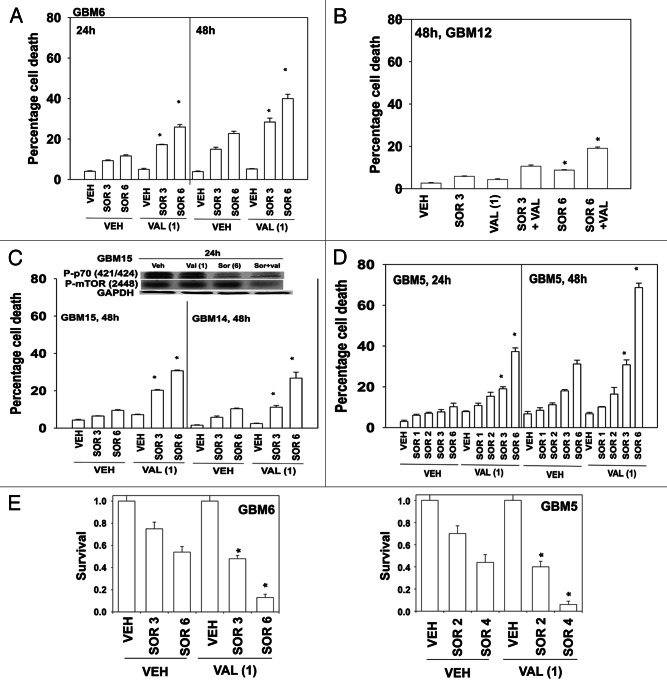
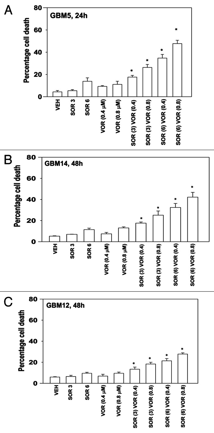
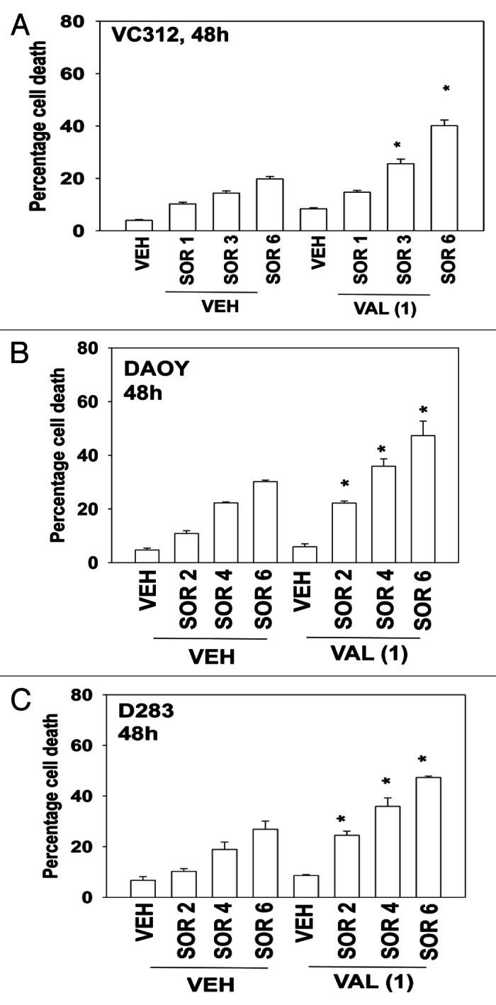
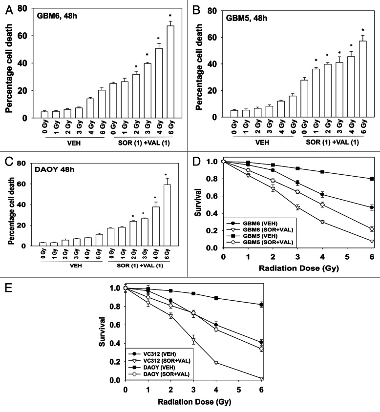
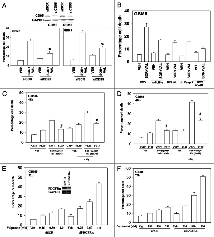
The present studies were designed to determine whether the multi-kinase inhibitor sorafenib (Nexavar) interacted with histone deacetylase inhibitors to kill glioblastoma and medulloblastoma cells. In a dose-dependent fashion sorafenib lethality was enhanced in multiple genetically disparate primary human glioblastoma isolates by the HDAC inhibitor sodium valproate (Depakote). Drug exposure reduced phosphorylation of p70 S6K and of mTOR. Similar data to that with valproate were also obtained using the HDAC inhibitor vorinostat (Zolinza). Sorafenib and valproate also interacted to kill medulloblastoma and PNET cell lines. Treatment with sorafenib and HDAC inhibitors radio-sensitized both GBM and medulloblastoma cell lines. Knock down of death receptor (CD95) expression protected GBM cells from the drug combination, as did overexpression of c-FLIP-s, BCL-XL and dominant negative caspase 9. Knock down of PDGFRα recapitulated the effect of sorafenib in combination with HDAC inhibitors. Collectively, our data demonstrate that the combination of sorafenib and HDAC inhibitors kills through activation of the extrinsic pathway, and could represent a useful approach to treat CNS-derived tumors.


Figures

None
Figure 1. Sorafenib and Sodium Valproate interact to kill GBM cells. (A) GBM6 cells were treated with vehicle or Sodium Valproate (1 mM). Thirty minutes later cells were treated with vehicle or sorafenib (3 μM; 6 μM). Cells were isolated 24 h and 48 h later. Viability was determined by trypan blue exclusion (n = 3, ± SEM) *p < 0.05 greater than corresponding vehicle control treated cells. (B) GBM12 cells were treated with vehicle or Sodium Valproate (1 mM). Thirty minutes later cells were treated with vehicle or sorafenib (3 μM; 6 μM). Cells were isolated 48 h later. Viability was determined by trypan blue exclusion (n = 3, ± SEM) *p < 0.05 greater than corresponding vehicle control treated cells. (C) GBM15 and GBM14 cells were treated with vehicle or Sodium Valproate (1 mM). Thirty minutes later cells were treated with vehicle or sorafenib (3 μM; 6 μM). Cells were isolated 48 h later. Viability was determined by trypan blue exclusion (n = 3, ± SEM) *p < 0.05 greater than corresponding vehicle control treated cells. Upper blot: GBM15 cells were treated with the indicated drugs and cells isolated 24 h later for determination of P-p70 S6K and P-mTOR levels. (D) GBM5 cells were treated with vehicle or Sodium Valproate (1 mM). Thirty minutes later cells were treated with vehicle or sorafenib (1 μM; 2 μM; 3 μM; 6 μM). Cells were isolated 24 h and 48 h later. Viability was determined by trypan blue exclusion (n = 3, ± SEM) *p < 0.05 greater than corresponding vehicle control treated cells. (E) GBM5 and GBM6 cells plated as single cells were treated with vehicle or Sodium Valproate (1 mM). Thirty minutes later cells were treated with vehicle or sorafenib (2 μM–6 μM, as shown). The media was changed 48 h later to drug free media and colonies permitted to form for the next 14 d. (n = 3, ± SEM) *p < 0.05 greater killing corresponding vehicle control treated cells.
None
Figure 2. Sorafenib and Vorinostat interact to kill GBM cells. (A) GBM5 cells were treated with vehicle or Vorinostat (0.4 μM; 0.8 μM). Thirty minutes later cells were treated with vehicle or sorafenib (3 μM; 6 μM). Cells were isolated 24 h later. Viability was determined by trypan blue exclusion (n = 3, ± SEM) *p < 0.05 greater than corresponding vehicle control treated cells. (B) GBM14 cells were treated with vehicle or Vorinostat (0.4 μM; 0.8 μM). Thirty minutes later cells were treated with vehicle or sorafenib (3 μM; 6 μM). Cells were isolated 24 h later. Viability was determined by trypan blue exclusion (n = 3, ± SEM) *p < 0.05 greater than corresponding vehicle control treated cells. (C) GBM12 cells were treated with vehicle or Vorinostat (0.4 μM; 0.8 μM). Thirty minutes later cells were treated with vehicle or sorafenib (3 μM; 6 μM). Cells were isolated 24 h later. Viability was determined by trypan blue exclusion (n = 3, ± SEM) *p < 0.05 greater than corresponding vehicle control treated cells.
None
Figure 3. Sorafenib and Vorinostat interact to kill medulloblastoma and PNET cells. (A) VC312 cells were treated with vehicle or Sodium Valproate (1 mM). Thirty minutes later cells were treated with vehicle or sorafenib (1 μM; 3 μM; 6 μM). Cells were isolated 48 h later. Viability was determined by trypan blue exclusion (n = 3, ± SEM) *p < 0.05 greater than corresponding vehicle control treated cells. (B) DAOY cells were treated with vehicle or Sodium Valproate (1 mM). Thirty minutes later cells were treated with vehicle or sorafenib (2 μM; 4 μM; 6 μM). Cells were isolated 48 h later. Viability was determined by trypan blue exclusion (n = 3, ± SEM) *p < 0.05 greater than corresponding vehicle control treated cells. (C) D283 cells were treated with vehicle or Sodium Valproate (1 mM). Thirty minutes later cells were treated with vehicle or sorafenib (2 μM; 4 μM; 6 μM). Cells were isolated 48 h later. Viability was determined by trypan blue exclusion (n = 3, ± SEM) *p < 0.05 greater than corresponding vehicle control treated cells.
None
Figure 4. Sorafenib and HDAC inhibitors radiosensitize GBM cells. (A) GBM6 cells were treated with vehicle or Sodium Valproate (1 mM). Thirty minutes later cells were treated with vehicle or sorafenib (1 μM). Thirty minutes later cells were exposed to ionizing radiation (0–6 Gy). Cells were isolated 48 h later. Viability was determined by trypan blue exclusion (n = 3, ± SEM) *p < 0.05 greater than corresponding vehicle control treated cells. (B) GBM5 cells were treated with vehicle or Sodium Valproate (1 mM). Thirty minutes later cells were treated with vehicle or sorafenib (1 μM). Thirty minutes later cells were exposed to ionizing radiation (0–6 Gy). Cells were isolated 48 h later. Viability was determined by trypan blue exclusion (n = 3, ± SEM) *p < 0.05 greater than corresponding vehicle control treated cells. (C) DAOY cells were treated with vehicle or Sodium Valproate (1 mM). Thirty minutes later cells were treated with vehicle or sorafenib (1 μM). Thirty minutes later cells were exposed to ionizing radiation (0–6 Gy). Cells were isolated 48 h later. Viability was determined by trypan blue exclusion (n = 3, ± SEM) *p < 0.05 greater than corresponding vehicle control treated cells. (D) GBM5 and GBM6 cells plated as single cells were treated with vehicle or Sodium Valproate (1 mM). Thirty minutes later cells were treated with vehicle or sorafenib (1 μM). Thirty minutes later cells were irradiated. The media was changed 48 h later to drug free media and colonies permitted to form for the next 14 d (n = 3, ± SEM). (E) DAOY and VC312 cells plated as single cells were treated with vehicle or Sodium Valproate (1 mM). Thirty minutes later cells were treated with vehicle or sorafenib (1 μM). Thirty minutes later cells were irradiated. The media was changed 48 h later to drug free media and colonies permitted to form for the next 14 d (n = 3, ± SEM).
None
Figure 5. Death receptor signaling is essential for sorafenib and valproate toxicity. (A) GBM5 and GBM6 cells were transfected with either scrambled control (siSCR) or an siRNA to knock down CD95 expression. Twenty-four h after transfection cells were treated with vehicle or valproate (1 mM) and sorafenib (3 μM). Cells were isolated 48 h later. Viability was determined by trypan blue exclusion (n = 3, ± SEM) #p < 0.05 less than corresponding vehicle control treated cells. (B) GBM5 cells were infected with the indicated recombinant adenoviruses (50 min.o.i.). Twenty-four h after infection as shown portions of cells were treated with the pan-caspase inhibitor zVAD (25 μM). Cells were isolated 48 h later. Viability was determined by trypan blue exclusion (n = 3, ± SEM). (C and D) GBM6 (C) and GBM5 (D) cells were infected with an empty vector virus or a virus to express c-FLIP-s. Twenty-four h after infection cells were treated with vehicle or valproate (1 mM) and sorafenib (3 μM). Thirty minutes later cells were irradiated (4 Gy). Cells were isolated 48 h later. Viability was determined by trypan blue exclusion (n = 3, ± SEM). (E and F) GBM5 cells were transfected with scrambled siRNA (siSCR) or an siRNA to knock down PDGFRα. Twenty-four h after transfection cells were treated with Sodium Valproate or Vorinostat. Cells were isolated 48 h later. Viability was determined by trypan blue exclusion (n = 3, ± SEM).

References

    1. Squatrito M, Holland EC. DNA damage response and growth factor signaling pathways in gliomagenesis and therapeutic resistance. Cancer Res. 2011;71:5945–9. doi: 10.1158/0008-5472.CAN-11-1245. - DOI - PubMed
    1. Buonerba C, Di Lorenzo G, Marinelli A, Federico P, Palmieri G, Imbimbo M, et al. A comprehensive outlook on intracerebral therapy of malignant gliomas. Crit Rev Oncol Hematol. 2011;80:54–68. doi: 10.1016/j.critrevonc.2010.09.001. - DOI - PubMed
    1. Bonavia R, Inda MM, Cavenee WK, Furnari FB. Heterogeneity maintenance in glioblastoma: a social network. Cancer Res. 2011;71:4055–60. doi: 10.1158/0008-5472.CAN-11-0153. - DOI - PMC - PubMed
    1. Liu KW, Hu B, Cheng SY. Platelet-derived growth factor receptor alpha in glioma: a bad seed. Chin J Cancer. 2011;30:590–602. doi: 10.5732/cjc.011.10236. - DOI - PMC - PubMed
    1. Park MA, Reinehr R, Häussinger D, Voelkel-Johnson C, Ogretmen B, Yacoub A, et al. Sorafenib activates CD95 and promotes autophagy and cell death via Src family kinases in gastrointestinal tumor cells. Mol Cancer Ther. 2010;9:2220–31. doi: 10.1158/1535-7163.MCT-10-0274. - DOI - PMC - PubMed

MeSH terms