

Cortical networks subserving upper limb movements in primates
- PMID: 22407009
- PMCID: PMC3695617
Cortical networks subserving upper limb movements in primates
Abstract
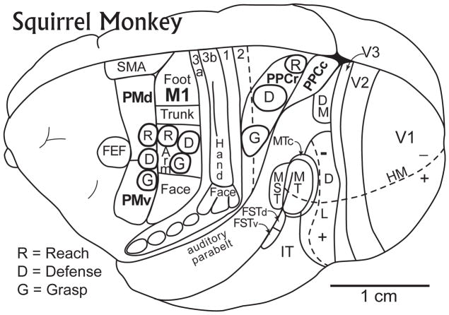
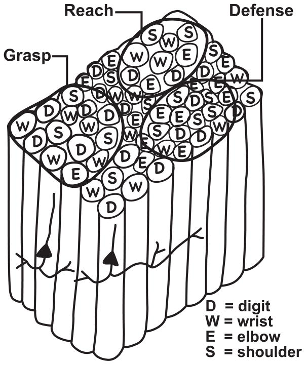
In all primates, the cortical control of hand and arm movements is initiated and controlled by a network of cortical regions including primary motor cortex (M1), premotor cortex (PMC), and posterior parietal cortex (PPC). These interconnected regions are influenced by inputs from especially visual and somatosensory cortical areas, and prefrontal cortex. Here we discuss recent evidence showing M1, PMC, and PPC can be subdivided into a number of functional zones or domains, including several that participate in guiding and controlling hand and arm movements. Functional zones can be defined by the movement sequences evoked by microstimulation within them, and functional zones related to the same type of movement in all three cortical regions are interconnected. The inactivation of a functional zone in each of the regions has a different impact on motor behavior. Finally, there is considerable plasticity within the networks so that behavioral recoveries can occur after damage to functional zones within a network.
Conflict of interest statement
Conflicts of interest: none
Figures
Similar articles
-
The dorsal stream of visual processing and action-specific domains in parietal and frontal cortex in primates.J Comp Neurol. 2023 Dec;531(18):1897-1908. doi: 10.1002/cne.25489. Epub 2023 Apr 28. J Comp Neurol. 2023. PMID: 37118872 Free PMC article. Review.
-
Effects of muscimol inactivations of functional domains in motor, premotor, and posterior parietal cortex on complex movements evoked by electrical stimulation.J Neurophysiol. 2014 Mar;111(5):1100-19. doi: 10.1152/jn.00491.2013. Epub 2013 Dec 18. J Neurophysiol. 2014. PMID: 24353298 Free PMC article.
-
Organization of the posterior parietal cortex in galagos: II. Ipsilateral cortical connections of physiologically identified zones within anterior sensorimotor region.J Comp Neurol. 2009 Dec 20;517(6):783-807. doi: 10.1002/cne.22190. J Comp Neurol. 2009. PMID: 19844952 Free PMC article.
-
Cortical connections of functional zones in posterior parietal cortex and frontal cortex motor regions in new world monkeys.Cereb Cortex. 2011 Sep;21(9):1981-2002. doi: 10.1093/cercor/bhq260. Epub 2011 Jan 24. Cereb Cortex. 2011. PMID: 21263034 Free PMC article.
-
Evolution of posterior parietal cortex and parietal-frontal networks for specific actions in primates.J Comp Neurol. 2016 Feb 15;524(3):595-608. doi: 10.1002/cne.23838. Epub 2015 Jul 21. J Comp Neurol. 2016. PMID: 26101180 Free PMC article. Review.
Cited by
-
Evidence for an effector-independent action system from people born without hands.Proc Natl Acad Sci U S A. 2020 Nov 10;117(45):28433-28441. doi: 10.1073/pnas.2017789117. Epub 2020 Oct 26. Proc Natl Acad Sci U S A. 2020. PMID: 33106395 Free PMC article.
-
A Web-Based Atlas Combining MRI and Histology of the Squirrel Monkey Brain.Neuroinformatics. 2019 Jan;17(1):131-145. doi: 10.1007/s12021-018-9391-z. Neuroinformatics. 2019. PMID: 30006920 Free PMC article.
-
Intracortical Microstimulation Maps of Motor, Somatosensory, and Posterior Parietal Cortex in Tree Shrews (Tupaia belangeri) Reveal Complex Movement Representations.Cereb Cortex. 2017 Feb 1;27(2):1439-1456. doi: 10.1093/cercor/bhv329. Cereb Cortex. 2017. PMID: 26759478 Free PMC article.
-
On the evolution of handedness: evidence for feeding biases.PLoS One. 2013 Nov 13;8(11):e78967. doi: 10.1371/journal.pone.0078967. eCollection 2013. PLoS One. 2013. PMID: 24236078 Free PMC article.
-
The dorsal stream of visual processing and action-specific domains in parietal and frontal cortex in primates.J Comp Neurol. 2023 Dec;531(18):1897-1908. doi: 10.1002/cne.25489. Epub 2023 Apr 28. J Comp Neurol. 2023. PMID: 37118872 Free PMC article. Review.
References
-
- Woolsey CN. In: Biological and Biochemical Bases of Behavior. Harlow HF, Woolsey CN, editors. U of Wisconsin Press; 1958. pp. 63–81.
-
- Schieber MH. Constraints on somatotopic organization in the primary motor cortex. J Neurophysiol. 2001;86:2125–2143. - PubMed
-
- Asanuma H, Rosen I. Topographic organization of cortical efferent zones projecting to distal forelimb muscles in the monkey. Exp Brain Res. 1972;14:243–256. - PubMed
-
- Leyton ASF, Sherrington CS. Observations on the excitable cortex of the chimpanzee, orangutan, and gorilla. Quart J Exp Physiol. 1917;11:137–222.
-
- Gould HJ, Cusick CG, Pons TP, Kaas JH. The relationship of corpus callosum connections to electrical stimulation maps of motor, supplementary motor, and the frontal eye fields in owl monkeys. J Comp Neurol. 1986;247:297–325. - PubMed
Publication types
MeSH terms
Grants and funding
LinkOut - more resources
Full Text Sources