. 2012 Mar 11;19(4):387-94.
doi: 10.1038/nsmb.2245.

A telomere-dependent DNA damage checkpoint induced by prolonged mitotic arrest

Affiliations

A telomere-dependent DNA damage checkpoint induced by prolonged mitotic arrest

Makoto T Hayashi et al. Nat Struct Mol Biol. .

Abstract

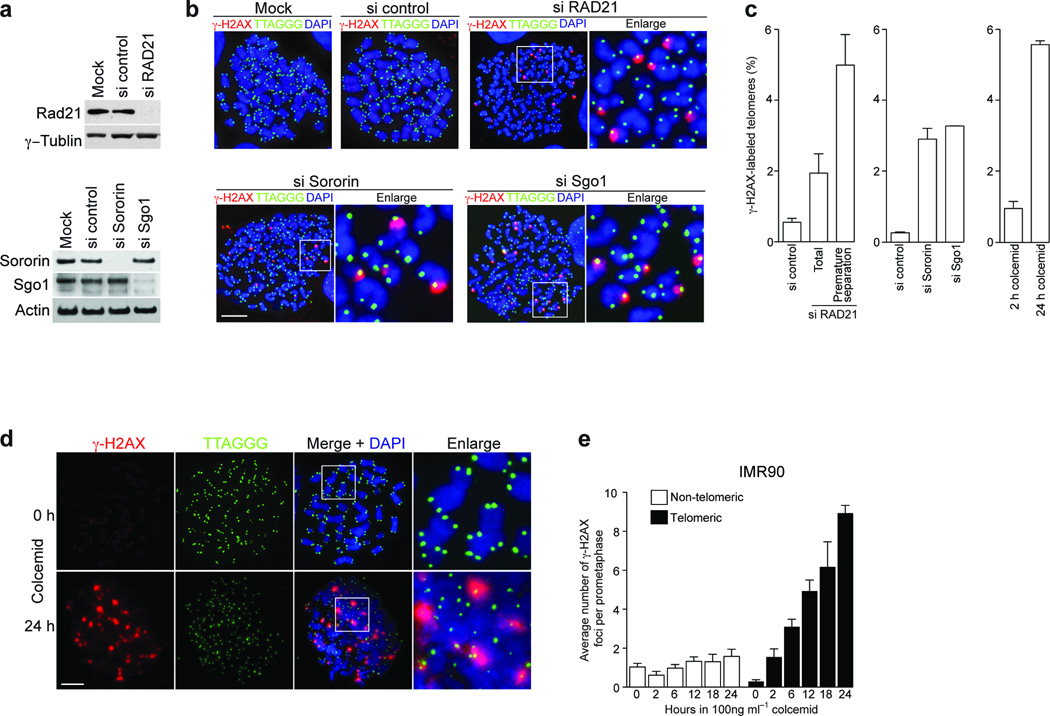
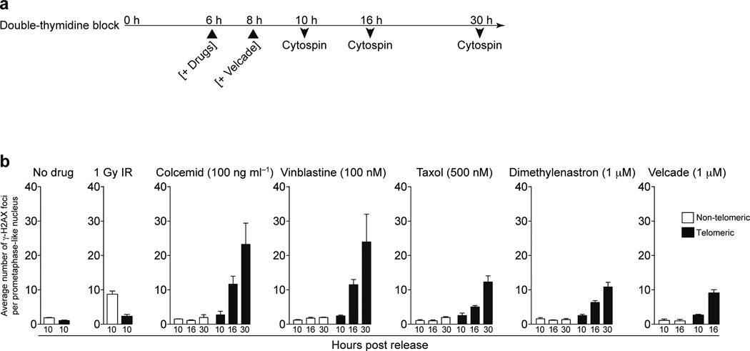
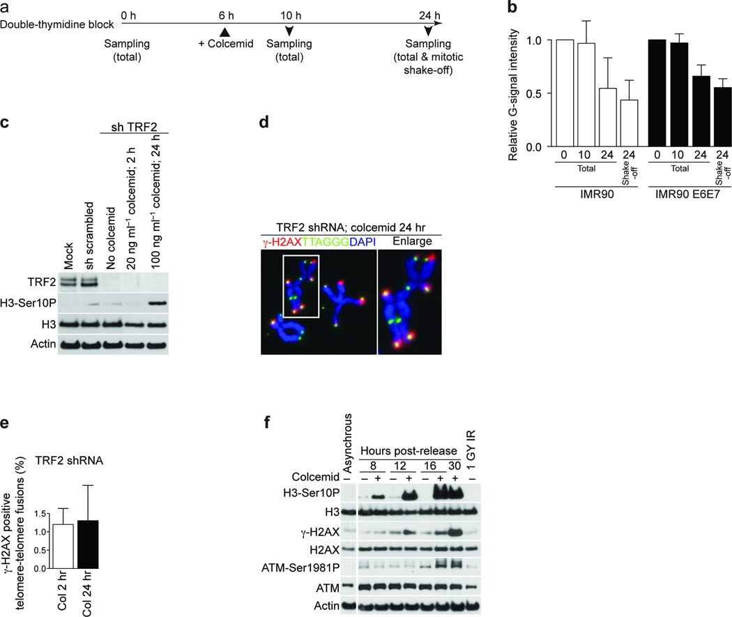
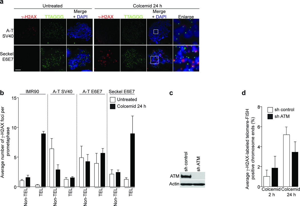
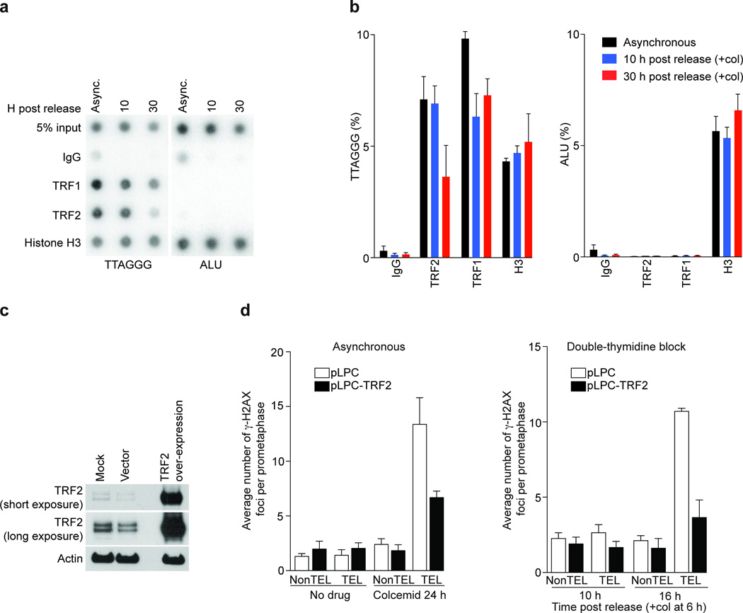
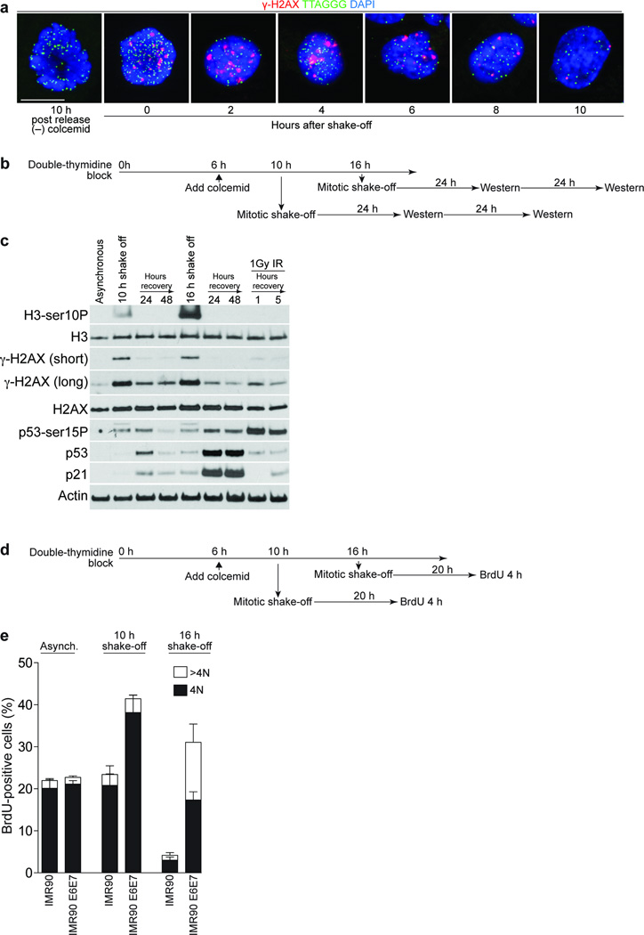
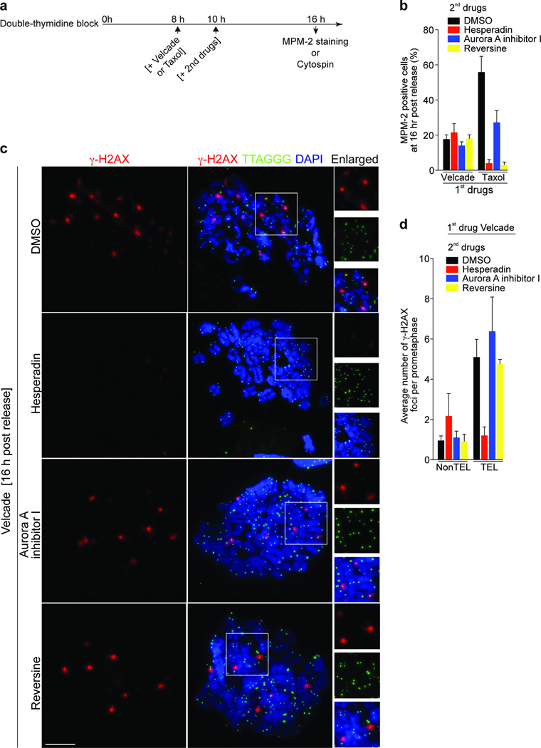
Telomere shortening and disruption of telomeric components are pathways that induce telomere deprotection. Here we describe another pathway, in which prolonged mitotic arrest induces damage signals at telomeres in human cells. Exposure to microtubule drugs, kinesin inhibitors, proteasome inhibitors or the disruption of proper chromosome cohesion resulted in the formation of damage foci at telomeres. Induction of mitotic telomere deprotection coincided with dissociation of TRF2 from telomeres, telomeric 3'-overhang degradation and ATM activation, and deprotection could be suppressed by TRF2 overexpression or inhibition of Aurora B kinase. Normal cells that escaped from prolonged mitotic arrest halted in the following G1 phase, whereas cells lacking p53 continued to cycle and became aneuploid. We propose a telomere-dependent mitotic-duration monitoring system that reacts to improper progression through mitosis.


Figures

Figure 1
Figure 1
Prolonged mitotic arrest causes telomere deprotection. (a) Western analysis of HeLa1.2.11 cells transfected with control siRNAs or siRNAs targeting RAD21, Sororin or Shugoshin (Sgo1). RAD21 has been targeted for 48 h, Sororin and Shugoshin for 24 h. γ-Tubulin and Actin serve as loading controls. (b) Immunofluorescence analysis of HeLa1.2.11 metaphase cells transfected with control siRNAs or siRNAs targeting RAD21, Sororin or Shugoshin. (c) Quantification of γ-H2AX positive telomeres in HeLa1.2.11 cells transfected as in panel a. The mean and standard deviation of three experiments quantifying 25 metaphases is shown. For RAD21 the percentages of γ-H2AX positive telomeres in all metaphases and metaphases that carry prematurely separated chromatids have been indicated, for Sororin and Shugoshin only the percentage in all metaphases is shown. γ-H2AX positive metaphase telomeres of HeLa1.2.11 cells treated with 100 ng ml−1 colcemid for 2 or 24 h are shown on the right. See also Supplementary Fig. 1a. (d) Immunofluorescence staining of metaphase chromosomes of IMR90 primary fibroblasts treated with colcemid for 0 hours or 24 hours. See also Supplementary Fig. 1b. (e) Quantification of γ-H2AX foci in IMR90 fibroblast prometaphases following the indicated treatment, displayed as in panel c. The mean and standard deviation of three experiments quantifying at least 25 prometaphases is shown. Scale bar, 10 µm.
Figure 2
Figure 2
Mitotic inhibitors cause telomere deprotection. (a) Schematic of the timing of experiments in panel b. (b) Quantification of γ-H2AX positive foci in prometaphase IMR90 cells following the indicated treatment. The mean and standard deviation of three experiments quantifying at least 25 prometaphases is shown.
Figure 3
Figure 3
Prolonged mitotic arrest causes the loss of 3’-overhangs and ATM activation. (a) Schematic of the timing of experiments in panel b. (b) Quantification of the ratio between native and denatured signals from telomeric southern analysis of IMR90 cells (white bars) or IMR90 E6E7 cells (black bars). The mean and standard deviation of three experiments is shown. (c) Western analysis of mock treated IMR90 E6E7 fibroblasts and cells expressing a scrambled shRNA or a shRNA targeting TRF2 with or without the indicated colcemid treatment. (d) Immunofluorescence image of metaphase chromosomes from IMR90 E6E7 cells with suppressed TRF2, exposed to 100 ng ml-1 colcemid for 24 hrs. (e) Quantification of γ-H2AX positive telomere fusions in IMR90 cells with suppressed TRF2, exposed to 20 ng ml-1 colcemid for 2 h or 100 ng ml-1 colcemid for 24 h. The mean and standard deviation of three experiments quantifying at least 135 fusions per experiment is shown. (f) Western analysis of asynchronous IMR90 fibroblasts and IMR90 cells released from G1-S arrest as shown. Where indicated, colcemid has been added 6 h after release.
Figure 4
Figure 4
Mitotic TIF formation is ATM dependent. (a) Immunofluorescence staining of A-T SV40 and Seckel E6E7 fibroblasts treated with colcemid for 24 h. Magnifications of indicated regions are on the right. (b) Quantification of γ-H2AX foci in IMR90, A-T SV40, A-T E6E7 and Seckel E6E7 metaphases. Displayed as in Fig. 1e. The mean and standard deviation of three independent experiments of 25 or more metaphases is shown. (c) Western analysis of ATM targeting in HeLa1.2.11 cells. (d) Quantification of meta-TIF in ATM targeted HeLa1.2.11 cells. Displayed as in Fig 1c. Scale bar, 10 µm.
Figure 5
Figure 5
Mitotic TIF are dependent on TRF2 removal. (a) Chromatin immunoprecipitation experiments from asynchronous IMR90 cells and synchronized IMR90 cells 10 and 30 h post release. Colcemid was added 6 h post release. Antibodies are indicated on the left and 5% of the input is shown. (b) Mean and standard error of three experiments as shown in panel a. (c) Western analysis of TRF2 overexpression in IMR90 cells. (d) Quantification of γ-H2AX foci in prometaphases from control and TRF2 overexpressing IMR90 cells in asynchronous (24 h of colcemid) and synchronized (10 and 16 h post release) populations. Displayed as in Fig. 1e.
Figure 6
Figure 6
Checkpoint activation by mitotic telomere deprotection. (a) Immunofluorescence of synchronized IMR90 cells treated with 100 ng ml-1 colcemid 6 h post release, cultured for 18 h before collection by shake-off. The cells were replated and fixated at the indicated times. A mitotic cell 10 h post-release without colcemid serves as the negative control. (b) Schematic of the timing of the experiment in panel c. (c) Western analysis of asynchronous IMR90 cells or IMR90 cells synchronized and collected as in panel b. Antibodies used are indicated on the left. (d) Schematic of the timing of the experiment in panel e. (e) Quantification of BrdU-positive cells in asynchronous IMR90 and synchronized IMR90 and IMR90 E6E7 populations treated as in panel d. BrdU incorporation and DNA content was determined by FACS analysis. The mean and standard deviation of three independent experiments (20,000 cells analyzed per time point) is shown. Scale bar, 10 µm.
Figure 7
Figure 7
Mitotic TIF formation is dependent on Aurora B kinase but not on the SAC. (a) Schematic of the timing of experiments in panels b-d. (b) Quantification of MPM-2 positive IMR90 cells that were first treated with 1µM Velcade or 500 nM Taxol, then control-treated with DMSO, with Hesperadin (250nM), Aurora A inhibitor I (3 µM) or Reversine (0.5 µM). (c) Meta-TIF analysis of IMR90 cells treated as indicated in panel d. Quantification of γ-H2AX foci from panel c. Displayed as in Fig. 1e. Scale bar, 10 µm.
Figure 8
Figure 8
Model for a telomere based mitotic duration checkpoint.

References

    1. de Lange T. How telomeres solve the end-protection problem. Science. 2009;326:948–952. - PMC - PubMed
    1. van Steensel B, Smogorzewska A, de Lange T. TRF2 protects human telomeres from end-to-end fusions. Cell. 1998;92:401–413. - PubMed
    1. Karlseder J, Broccoli D, Dai Y, Hardy S, de Lange T. p53- and ATM-dependent apoptosis induced by telomeres lacking TRF2. Science. 1999;283:1321–1325. - PubMed
    1. Denchi EL, de Lange T. Protection of telomeres through independent control of ATM and ATR by TRF2 and POT1. Nature. 2007;448:1068–1071. - PubMed
    1. Guo X, et al. Dysfunctional telomeres activate an ATM-ATR-dependent DNA damage response to suppress tumorigenesis. EMBO J. 2007;26:4709–4719. - PMC - PubMed

Publication types

MeSH terms