. 2012 Apr 18;31(8):2013-23.
doi: 10.1038/emboj.2012.55. Epub 2012 Mar 9.

Codanin-1, mutated in the anaemic disease CDAI, regulates Asf1 function in S-phase histone supply

Affiliations

Codanin-1, mutated in the anaemic disease CDAI, regulates Asf1 function in S-phase histone supply

Katrine Ask et al. EMBO J. .

Erratum in

  • EMBO J. 2012 Jul 18;31(14):3229

Abstract

Efficient supply of new histones during DNA replication is critical to restore chromatin organization and maintain genome function. The histone chaperone anti-silencing function 1 (Asf1) serves a key function in providing H3.1-H4 to CAF-1 for replication-coupled nucleosome assembly. We identify Codanin-1 as a novel interaction partner of Asf1 regulating S-phase histone supply. Mutations in Codanin-1 can cause congenital dyserythropoietic anaemia type I (CDAI), characterized by chromatin abnormalities in bone marrow erythroblasts. Codanin-1 is part of a cytosolic Asf1-H3.1-H4-Importin-4 complex and binds directly to Asf1 via a conserved B-domain, implying a mutually exclusive interaction with the chaperones CAF-1 and HIRA. Codanin-1 depletion accelerates the rate of DNA replication and increases the level of chromatin-bound Asf1, suggesting that Codanin-1 guards a limiting step in chromatin replication. Consistently, ectopic Codanin-1 expression arrests S-phase progression by sequestering Asf1 in the cytoplasm, blocking histone delivery. We propose that Codanin-1 acts as a negative regulator of Asf1 function in chromatin assembly. This function is compromised by two CDAI mutations that impair complex formation with Asf1, providing insight into the molecular basis for CDAI disease.


Conflict of interest statement

The authors declare that they have no conflict of interest.

Figures

Figure 1
Figure 1
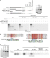
Codanin-1, a new partner of the cytosolic Asf1–H3-H4–Importin-4 complex. (A) Coomassie staining of Asf1a complexes isolated from asynchronous HeLa S3 cells stably expressing Onestrep-tagged (e−) Asf1a. Codanin-1 was identified in both cytosolic and nuclear e-Asf1a complexes by mass spectrometry analysis. Proteins annotated in black were previously reported (Groth et al, 2007a; Jasencakova et al, 2010). (B) Co-immunoprecipitation of Codanin-1 with endogenous Asf1 (a and b) from cytosolic HeLa S3 extracts. (C) Size-exclusion chromatography of cytosolic e-Asf1b complexes isolated as in (A). Codanin-1 co-elutes together with histone H3 and Importin-4. (D) Co-immunoprecipitation of Importin-4 and Asf1 (a and b) with endogenous Codanin-1 from cytosolic HeLa S3 extracts. (E) FLAG–HA-tagged (e−) H3.1 was immunoprecipitated from cytosolic extracts of HeLa S3 cells expressing e-H3.1 (Tagami et al, 2004) and analysed by western blotting. Pull down with sepharose beads was used as negative control. (F) Western blot analysis of cytosolic complexes containing wild-type e-Asf1a or the histone binding mutant, e-Asf1a V94R.
Figure 2
Figure 2
Codanin-1 binds directly to Asf1 via the same pocket as HIRA and CAF-1. (A) (Top left) Summary of the in vitro analysis of Codanin-1–Asf1 binding. ‘+’, binding; ‘−’, no binding; n.d., not done. (Top right) Coomassie staining of GST–Asf1 used for the pull-down assay. (Lower panel) In vitro binding analysis using GST–Asf1 (a and b) to pull down in vitro translated 35S-labelled Codanin-1. Bound proteins were visualized by autoradiography. (B) (Left) Sequence alignment of the B-domains in Codanin-1, HIRA and CAF-1 p60. Note that CAF-1 p60 has two B-domain-like motifs designated (1) and (2). Conservation is indicated by red colour intensity. (Right) Sequence alignment showing conservation of the Codanin-1 B-domain in human (Homo sapiens, H.s.), mice (Mus musculus, M.m.), opossum (Monodelphis domestica, M.d.), frogs (Xenopus tropicalis, X.t.), fish (Danio rerio, D.r.) and flies (Drosophila melanogaster, D.m.). The degree of conservation is illustrated by red colour intensity. (C) (Left) Coomassie staining of GST–Asf1 used for the pull-down assay. (Right) In vitro binding analysis using GST fusions of Asf1a, Asf1b and Asf1b mutated in the B-domain binding pocket (Asf1b D36AD37A; Tang et al, 2006) and in vitro translated 35S-labelled wild-type Codanin-1 or B-domain mutant (SRR/AAA). Bound proteins were visualized by autoradiography.
Figure 3
Figure 3
Codanin-1 depletion enhances DNA replication and Asf1 binding to chromatin. (A) U-2-OS cells were treated with an independent siRNA (siCdan1 #1) or an siRNA smart pool (siCdan1 #2) targeting Codanin-1 for 56 or 70 h. Knockdown efficiency was assessed by western blot analysis and qPCR (Supplementary Figure S3A). (B) Immunofluorescence analysis of U-2-OS cells treated with siRNA for 56 h followed by EdU pulse labelling. PCNA staining served as a marker for S-phase cells. Scale bar, 20 μm. (C) Quantification of EdU incorporation. (Left) Dot plot illustrating the distribution of EdU intensities within one experiment. Cells were treated as in (B) and EdU intensities were measured in PCNA-positive cells. n>87 and ***P<0.0001 calculated by Wilcoxon paired test. (Right) Bar diagram showing the average of three independent experiments with error bars indicating standard deviation. The values of siControl were set to 100%. In each experiment, we analysed between 87 and 187 cells per sample. (D) Quantification of chromatin-bound PCNA in cells treated as in (B). The bar diagram shows the average of three independent experiments with error bars indicating standard deviation. The values of siControl were set to 100%. In each experiment, we analysed between 87 and 187 cells per sample. The distribution of PCNA levels within one experiment is shown in Supplementary Figure S4C. (E) Immunofluorescence analysis of chromatin-bound Asf1 in U-2-OS cells stably expressing RFP-PCNA to mark S-phase cells. Codanin-1 was depleted by RNAi for 56 h prior to pre-extraction of soluble proteins and fixation. Scale bar, 20 μm. (F) Quantification of chromatin-bound Asf1 in cells treated as in (E). Dot plot illustrating the distribution of nuclear Asf1a intensities in pre-extracted cells treated for 56 h with the indicated siRNAs. n>97 and ***P<0.0001 calculated by Wilcoxon paired test. This result is representative of three independent experiments and similar results were observed with an independent siRNA (Supplementary Figure S4D).
Figure 4
Figure 4
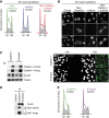
Ectopic Codanin-1 expression arrests S-phase progression by sequestering Asf1 in the cytoplasm. (A) Cell-cycle profiles of cells transfected with Myc–FLAG–Codanin-1 and/or e-Asf1a analysed 30 h post transfection. Cells were co-transfected with a GFP-Spectrin expression vector to gate for transfected cells. (B) Immunofluorescence analysis of cells treated as in (A). Cells were harvested 24 h after transfection and stained with antibodies against the Myc epitope and Asf1a. Scale bar, 20 μm. (C) Western blotting (right) and immunofluorescence analysis (left) of cells expressing ectopic wild-type FLAG–HA–Codanin-1 or a B-domain mutant. Conditional T-REx Flp-In U-2-OS cells were left untreated (−Tet) or induced with tetracycline for 24 h (+Tet). Scale bar, 20 μm. (D) Wild-type FLAG–HA-tagged Codanin-1 and B-mutant were immunoprecipitated from soluble protein extracts prepared from cells treated as in (C), and analysed by western blotting. (E) Cell-cycle profiles of cells from (C) analysed by FACS.
Figure 5
Figure 5
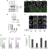
CDAI missense mutations disable the functional interaction with Asf1. (A) Western blotting (left) and immunofluorescence analysis (right) of cells expressing ectopic wild-type FLAG–HA–Codanin-1 or CDAI mutants R714W and R1042W. U-2-OS cells conditional for expression of the indicated Codanin-1 proteins were induced with tetracycline for 24 h. The parental U-2-OS Flp-In cell line was used as a negative control. Scale bar, 20 μm. (B) Wild-type FLAG–HA-tagged Codanin-1 and CDAI mutants R714W and R1042W were immunoprecipitated from soluble protein extracts prepared from cells treated as in (A) and analysed by western blotting (top). Quantitative determination of Asf1 binding to CDAI mutants (bottom). Asf1 binding was analysed by western blotting and quantified in three independent experiments. The graph shows Asf1 (a or b) signals normalized to FLAG with error bars indicating standard deviation. (C) Immunofluorescence analysis of Asf1 localization in cells treated as in (A). Merged panel shows DAPI in blue, Asf1a in red and HA signals in green. Images representative of three independent experiments are shown with the percentage of cells showing nuclear Asf1 staining indicated below. Scale bar, 10 μm. (D) Cell-cycle profiles of conditional cells induced as in (A) and compared with the non-induced parental cell line. (E) EdU incorporation in cells expressing wild-type FLAG–HA–Codanin-1 and CDAI mutants R714W and R1042W. EdU intensities were quantified as in Figure 3C. The mean intensity of non-induced parental cells was set to 100%. The average of three independent experiments is presented with error bars indicating standard deviation. In each experiment, we analysed between 70 and 204 cells per sample.
Figure 6
Figure 6
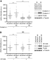
The R714W CDAI disease mutant cannot substitute for endogenous Codanin-1 in regulation of Asf1. Quantitative analysis of Asf1 binding to chromatin (left) and western blotting (right) in cells depleted for endogenous Codanin-1 followed by expression of ectopic FLAG–HA–Codanin-1 wild-type (A) or R714W mutant (B). Conditional cells (described in Figure 5A) were treated with siRNA to deplete endogenous Codanin-1 (siCdan1 #2) or control siRNAs (siControl) and 32 h later either induced (+) or left uninduced (−). After additional 24 h, cells were harvested for immunofluorescence analysis (left) and western blotting (right). Soluble proteins were removed by pre-extraction prior to fixation and cells were stained with antibodies against Asf1 and PCNA. Asf1 levels on chromatin were measured as in Figure 3E. n>86; ***P<0.0001; NS, not significant P=0.66 calculated by Wilcoxon paired test.
Figure 7
Figure 7
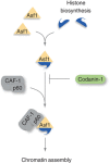
Model. We propose that Codanin-1 acts as a negative regulator of Asf1 function in provision of histones to replicating chromatin. Codanin-1 interacts directly with Asf1 and can sequester the chaperone in the cytoplasm away from replicating DNA. Moreover, Codanin-1 binding to Asf1 is mutually exclusive with CAF-1 and HIRA, making it a potential inhibitor of Asf1 histone donor function. In absence of Codanin-1, more Asf1 is present on chromatin, perhaps because Asf1–H3-H4 shuttling to the nucleus is accelerated. Concomitantly, cells show an increased rate of DNA synthesis, suggesting that Codanin-1 guards a limiting step in chromatin replication. We propose that Codanin-1 could regulate nuclear import of histones through its ability to bind the Asf1–H3.1-H4–Importin-4 complex. Only once Codanin-1 dissociates from the Asf1–H3.1-H4 complex in the nucleus, Asf1 would be able to bind CAF-1 and deliver histones for chromatin assembly.

References

    1. Alvarez F, Muñoz F, Schilcher P, Imhof A, Almouzni G, Loyola A (2011) Sequential establishment of marks on soluble histones H3 and H4. J Biol Chem 286: 17714–17721 - PMC - PubMed
    1. Annunziato AT (2011) Assembling chromatin: the long and winding road. Biochim Biophys Acta 1819: 196–210 - PubMed
    1. Campos EI, Fillingham J, Li G, Zheng H, Voigt P, Kuo W-HW, Seepany H, Gao Z, Day LA, Greenblatt JF, Reinberg D (2010) The program for processing newly synthesized histones H3.1 and H4. Nat Struct Mol Biol 17: 1343–1351 - PMC - PubMed
    1. Choudhary C, Olsen JV, Brandts C, Cox J, Reddy PN, Böhmer FD, Gerke V, Schmidt-Arras DE, Berdel WE, Müller-Tidow C, Mann M, Serve H (2009) Mislocalized activation of oncogenic RTKs switches downstream signaling outcomes. Mol Cell 36: 326–339 - PubMed
    1. Cook AJ, Gurard-Levin ZA, Vassias I, Almouzni G (2011) A specific function for the histone chaperone NASP to fine-tune a reservoir of soluble H3-H4 in the histone supply chain. Mol Cell 44: 918–927 - PubMed

Publication types

MeSH terms