. 2012 Mar 11;483(7391):627-31.
doi: 10.1038/nature10893.

Tissue factor and PAR1 promote microbiota-induced intestinal vascular remodelling

Affiliations

Tissue factor and PAR1 promote microbiota-induced intestinal vascular remodelling

Christoph Reinhardt et al. Nature. .

Abstract

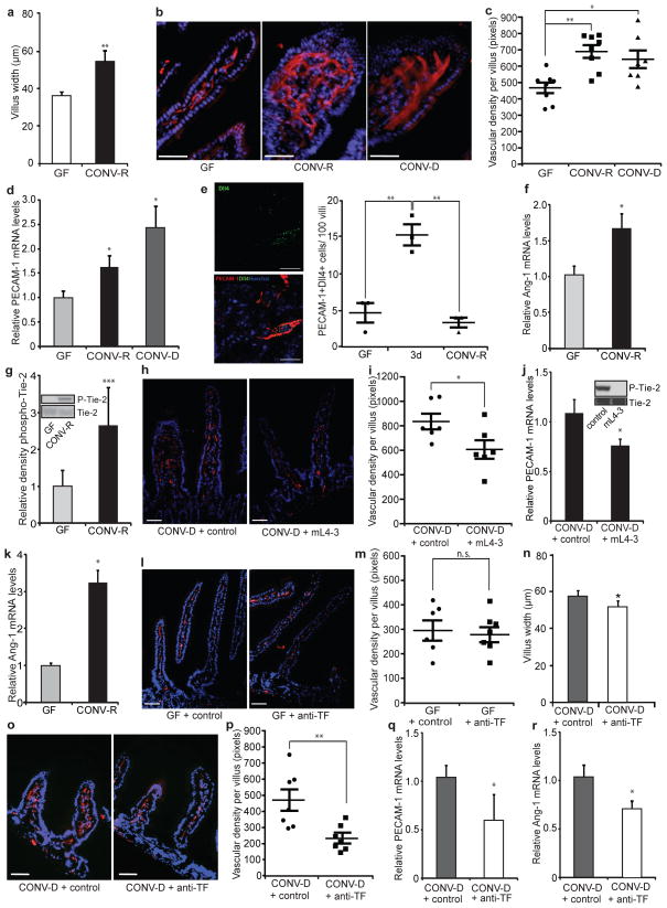
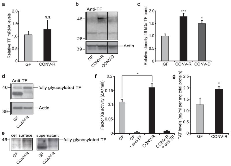
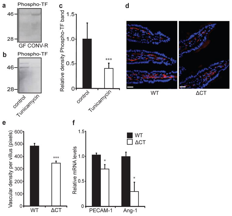
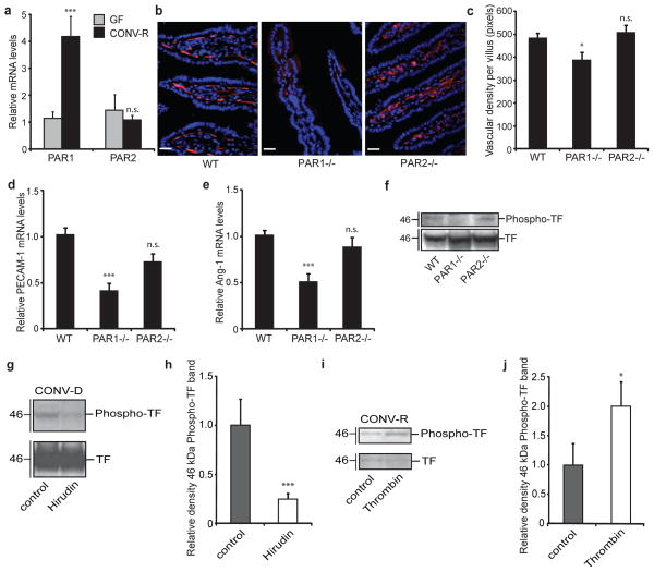
The gut microbiota is a complex ecosystem that has coevolved with host physiology. Colonization of germ-free (GF) mice with a microbiota promotes increased vessel density in the small intestine, but little is known about the mechanisms involved. Tissue factor (TF) is the membrane receptor that initiates the extrinsic coagulation pathway, and it promotes developmental and tumour angiogenesis. Here we show that the gut microbiota promotes TF glycosylation associated with localization of TF on the cell surface, the activation of coagulation proteases, and phosphorylation of the TF cytoplasmic domain in the small intestine. Anti-TF treatment of colonized GF mice decreased microbiota-induced vascular remodelling and expression of the proangiogenic factor angiopoietin-1 (Ang-1) in the small intestine. Mice with a genetic deletion of the TF cytoplasmic domain or with hypomorphic TF (F3) alleles had a decreased intestinal vessel density. Coagulation proteases downstream of TF activate protease-activated receptor (PAR) signalling implicated in angiogenesis. Vessel density and phosphorylation of the cytoplasmic domain of TF were decreased in small intestine from PAR1-deficient (F2r(-/-)) but not PAR2-deficient (F2rl1(-/-)) mice, and inhibition of thrombin showed that thrombin-PAR1 signalling was upstream of TF phosphorylation. Thus, the microbiota-induced extravascular TF-PAR1 signalling loop is a novel pathway that may be modulated to influence vascular remodelling in the small intestine.


Conflict of interest statement

The authors declare no competing financial interests.

Figures

Figure 1
Figure 1. TF promotes microbe-induced vascular remodelling in the gut
a, Villus width of sections of small intestine from GF and CONV-R mice (n = 4 mice per group). b, PECAM-1 staining (red) of sections of small intestine from GF, CONV-R and CONV-D mice. Nuclei were stained with Hoechst nuclear dye (blue). c, Quantification of b (n = 7 or 8 mice per group). d, Relative levels of mRNA for the vascular marker PECAM-1 in GF, CONV-R and CONV-D mice (n = 6 or 7 mice per group). e, Dll4 staining (green) of sections of small intestine from GF, ex-GF mice colonized for 3 days (3d), and CONV-R mice. Endothelial cells were stained with PECAM-1 (red). Dll4-positive endothelial cells per 100 villi were quantified (n = 3 mice per group). f, Relative levels of mRNA for Ang-1 in sections of small intestine from GF and CONV-R mice (n = 7–11 mice per group). g, Anti-phospho-Tie-2 immunoblot (Y1100 phosphorylation site) and quantification relative to total Tie-2 of small-intestinal lysates from GF and CONV-R mice (n = 5 mice per group). h, PECAM-1 staining of sections of small intestine from mice treated with control NaCl solution or the Ang-1 neutralizing peptibody mL4-3. i, Quantification of h (n = 6 mice per group). j, Relative levels of mRNA for PECAM-1 in sections of small intestine from CONV-D mice treated with NaCl control or mL4-3 (n = 10 or 11 mice per group). The inset shows that mL4-3 is a potent inhibitor of Ang-1-mediated Tie-2 phosphorylation. k, Relative levels of mRNA for Ang-1 in primary enterocytes from GF and CONV-R mice (n = 10 or 11 mice per group). l, PECAM-1 staining of sections of small intestine from GF mice treated with control or anti-TF antibody. m, Quantification of l (n = 6 or 7 mice per group). n, Villus width of sections of small intestine from CONV-D mice treated with control or anti-TF antibody (n = 4 mice per group). o, PECAM-1 staining of sections of small intestine from CONV-D mice treated with control or anti-TF antibody. p, Quantification of o (n = 7 mice per group). q, r, Relative levels of mRNA for PECAM-1 (q) and Ang-1 (r) in small intestine from CONV-D mice treated with control or anti-TF antibody (n = 5 or 6 mice per group). Female Swiss Webster mice or cells isolated from these mice were analysed in all panels. Scale bars, 50 μm. Results are shown as means ± s.e.m. Asterisk, P < 0.05; two asterisks, P < 0.01; three asterisks, P < 0.005; n.s., not significant.
Figure 2
Figure 2. The gut microbiota increases TF procoagulant activity and cell-surface localization
a, Relative levels of mRNA for TF in sections of small intestine from GF and CONV-R mice (n = 7–11 mice per group). b, Anti-TF immunoblot of small-intestinal lysates from GF, CONV-R and CONV-D mice. c, Quantification of the 46-kDa TF band shown in b (n = 14–25 mice per group). Data are normalized to actin and expressed relative to GF. d, Anti-TF immunoblot of primary enterocytes (from GF and CONV-R mice) after 2 h of culture. e, Anti-TF immunoblots from N-hydroxysuccinimido-biotin-labelled primary enterocytes from GF and CONV-R mice. Left: pull-down of proteins located on the plasma membrane with NeutrAvidin beads. Right: supernatant containing unlabelled proteins. f, Factor Xa activity in small-intestinal lysates from GF and CONV-R mice treated with control or anti-TF antibody (n = 4 or 5 mice per group). g, Levels of thrombin–antithrombin (TAT) complexes in small-intestinal lysates from GF and CONV-R mice (n = 7 mice per group). Female Swiss Webster mice or cells isolated from these mice were analysed in all panels. Results are shown as means ± s.e.m. Asterisk, P < 0.05; three asterisks, P < 0.005; n.s., not significant.
Figure 3
Figure 3. The gut microbiota increases phosphorylation of the cytoplasmic tail of TF, which increases vessel density in the intestine
a, b, Anti-phospho-TF immunoblot of (a) small-intestinal lysates from GF and CONV-R mice and (b) primary enterocytes (from CONV-R mice) incubated for 2 h in the absence and presence of tunicamycin (10 μmol l−1). c, Quantification of b (n = 5 mice per group). d, PECAM-1 staining (red) of sections of small intestine from 10–12-week-old wild-type (WT) and ΔCT female mice on a C57Bl6/J genetic background. Nuclei were stained with Hoechst nuclear dye (blue). e, Quantification of d (n = 4–6 mice per group). f, Relative levels of mRNA for PECAM-1 and Ang-1 in segments of small intestine from WT and ΔCT mice (n = 3 or 4 mice per group). Female Swiss Webster mice or cells isolated from these mice were analysed in a–c. Scale bars, 20 μm. Results are shown as means ± s.e.m. Asterisk, P < 0.05; three asterisks, P < 0.005.
Figure 4
Figure 4. PAR1 activation increases vessel density in the small intestine
a, Relative levels of mRNA for PAR1 and PAR2 in segments of small intestine from GF and CONV-R mice (n = 7 or 8 mice per group). b, PECAM-1 staining (red) of sections of small intestine from wild-type (WT), F2r−/− and F2rl1−/− mice. Nuclei were stained with Hoechst nuclear dye (blue). c, Quantification of b (n = 6–9 mice per group). d, e, Relative levels of mRNA for PECAM-1 (d) and Ang-1 (e) in segments of small intestine from wild-type, F2r−/− and F2rl1−/− mice (n = 6–9 mice per group). f, g, Anti-TF and anti-phospho-TF immunoblots of small-intestinal lysates from (f) WT, F2r−/− and F2rl1−/− miceand (g) CONV-D mice treated with PBS (control) or hirudin (1 mg/mouse) immediately before colonization and at 2 h and 4 h after colonization. h, Quantification of the phospho-TF band shown in g (n = 6 or 7 mice per group). i, Anti-TF and anti-phospho-TF immunoblots of primary enterocytes (from CONV-R mice) incubated for 2 h with human thrombin (50 nmol l−1). j, Quantification of the phospho-TF band shown in i (n = 8 mice per group). Female Swiss Webster mice were analysed in a and g–j. Female WT, F2r−/− and F2rl1−/− mice on a C57BL6/J genetic background were used in b–f. Scale bars, 20 μm. Results are shown as means ± s.e.m. Asterisk, P < 0.05; three asterisks, P < 0.005; n.s., not significant.

References

    1. Stappenbeck TS, Hooper LV, Gordon JI. Developmental regulation of intestinal angiogenesis by indigenous microbes via Paneth cells. Proc Natl Acad Sci USA. 2002;99:15451–15455. - PMC - PubMed
    1. Morrissey JH, Fakhrai H, Edgington TS. Molecular cloning of the cDNA for tissue factor, the cellular receptor for the initiation of the coagulation protease cascade. Cell. 1987;50:129–135. - PubMed
    1. Carmeliet P, et al. Role of tissue factor in embryonic blood vessel development. Nature. 1996;383:73–75. - PubMed
    1. Belting M, et al. Regulation of angiogenesis by tissue factor cytoplasmic domain signaling. Nature Med. 2004;10:502–509. - PubMed
    1. Griffin TC, Srinivasan Y, Zheng YW, Huang W, Coughlin SR. A role for thrombin receptor signaling in endothelial cells during embryonic development. Science. 2001;293:1666–1670. - PubMed

Publication types

MeSH terms