. 2012 Jun;15(2):287-303.
doi: 10.1007/s10456-012-9262-4. Epub 2012 Mar 11.

Calpain-mediated vimentin cleavage occurs upstream of MT1-MMP membrane translocation to facilitate endothelial sprout initiation

Affiliations

Calpain-mediated vimentin cleavage occurs upstream of MT1-MMP membrane translocation to facilitate endothelial sprout initiation

Hyeong-Il Kwak et al. Angiogenesis. 2012 Jun.

Abstract

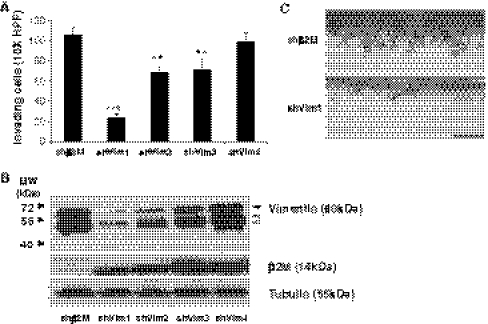
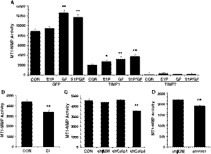
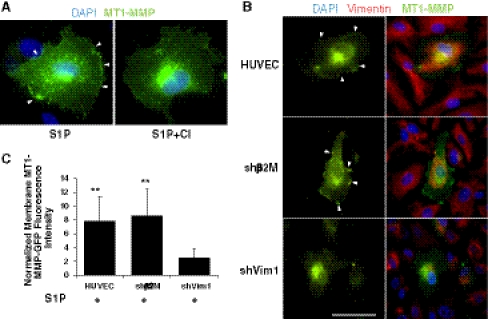
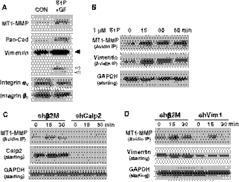
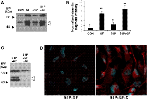
Endothelial cells normally line the vasculature and remain quiescent. However, these cells can be rapidly stimulated to undergo morphogenesis and initiate new blood vessel formation given the proper cues. This study reports a new mechanism for initiating angiogenic sprout formation that involves vimentin, the major intermediate filament protein in endothelial cells. Initial studies confirmed vimentin was required for sphingosine 1-phosphate (S1P)- and growth factor (GF)-induced endothelial cell invasion, and vimentin was cleaved by calpains during invasion. Calpains were predominantly activated by GF and were required for sprout initiation. Because others have reported membrane type 1-matrix metalloproteinase (MT1-MMP) is required for endothelial sprouting responses, we tested whether vimentin and calpain acted upstream of MT1-MMP. Both calpain and vimentin were required for successful MT1-MMP membrane translocation, which was stimulated by S1P. In addition, vimentin complexed with MT1-MMP in a manner that required both the cytoplasmic domain of MT1-MMP and calpain activation, which increased the soluble pool of vimentin in endothelial cells. Altogether, these data indicate that pro-angiogenic signals converge to activate calpain-dependent vimentin cleavage and increase vimentin solubility, which act upstream to facilitate MT1-MMP membrane translocation, resulting in successful endothelial sprout formation in three-dimensional collagen matrices. These findings help explain why S1P and GF synergize to stimulate robust sprouting in 3D collagen matrices.


Figures

Fig. 1
Fig. 1
Vimentin knockdown interfered with invasion responses. a Quantification of invasion density resulting from shRNA-mediated knockdown of beta 2 microglobulin (shβ2M) or vimentin (shVim). Four independent sequences are shown for vimentin knockdown (shVim1-4). Cells expressing indicated shRNA sequences were allowed to invade in the presence of S1P, VEGF, and bFGF, as previously described [32]. Cultures were fixed at 16 h of invasion and a representative experiment (n = 4) is shown. Data presented are average values from five 1 mm2 fields (±SD). ***P < 0.001, **P < 0.01 versus shβ2M, Student’s t test. b Western blot analyses of whole cell lysates of invading cells (16 h) using vimentin-, beta 2 microglobulin (β2M)-, and tubulin-specific antisera. Open arrowheads indicate vimentin cleavage products. c Representative photographs from a side view illustrate invasion responses observed with control and vimentin knockdown cells. Scale bar, 100 μm
Fig. 2
Fig. 2
Calpains are activated by growth factors and result in vimentin cleavage. a Invading cultures were treated with indicated doses of CI and allowed to invade collagen matrices for 22 h in the presence of S1P and GF. Whole cell extracts were subjected to Western blotting and probed with antisera directed to vimentin. Blots were stripped and re-probed with GAPDH- specific antisera. Results are representative of three independent experiments. b ECs were plated at 80% confluence in a 96 well plate in M199 medium containing RSII for 8 h. The cells were pre-treated with 31.6 μM CI or DMSO (CON) for 1 h and then loaded with 30 μM of the calpain substrate tBoc-LM-CMAC. Cells were treated without (CON) or with S1P (1 μM), GF (40 ng/ml VEGF and bFGF) or S1P + GF for 30 min and imaged. Calpain activity was quantified as indicated in the “Materials and methods” section. Results are representative of four independent experiments. Data shown are average values ± SD. *P < 0.05, and **P < 0.01 compared to Control, ***P < 0.001 compared to S1P + GF by Student’s t test. c Endothelial cells were seeded on 3D collagen matrices and allowed to invade for 4 or 6 h. Whole cell lysates were prepared and analyzed by Western blotting with antisera directed to vimentin and GAPDH control. d Quantification of intensities of vimentin cleavage products with treatment conditions. Data are derived by averaging band intensities from three independent experiments. **P < 0.01 versus CON; P < 0.05 versus all other treatments by Student’s t test
Fig. 3
Fig. 3
Endothelial cell invasion stimulated by S1P and GF requires calpain activation. a Invasion experiments were established by pre-incubating cells with CI at the concentrations indicated for 30 min prior to seeding on collagen matrices. Cells were allowed to invade for 22 h. Data represent average numbers of invading cells per standardized field ± SD (n = 4). b Representative photographs of a side view of invading cells from control (CON) and 100 μM CI treatment. Arrowhead indicates original monolayer. Black arrows indicate a lumen and white arrows indicate altered structures observed with CI treatment. c Quantification of average invasion distances (in microns) from monolayer to the tip of invading structures control (CON) and CI treated group (n = 100 cells). d Quantification of lumen diameter (in microns) of invading structures in control (CON) and 100 μM CI treated group (n = 100 cells). Results are representative of three independent experiments. Data shown in a, c, and d are average values ± SD. *P < 0.05, **P < 0.01 versus control by Student’s t test
Fig. 4
Fig. 4
Calpain knockdown significantly reduced EC invasion. ECs were not treated (CON) or transduced with lentiviruses delivering shRNA directed to beta 2 microglobulin (shβ2M; negative control), calpain 1 (shCalp1) and calpain 2 (shCalp2). Stable cell lines were selected with puromycin (0.2 μg/ml) for 2 weeks prior to testing in invasion assays. a Representative photographs of a side view of invading cells from each treatment group. Bar = 100 μm. b Quantification of the average number of invading cells per 0.25 mm2 field (n = 4 fields) from a representative experiment (n = 4); **P < 0.01 versus control, Student’s t test. c Quantification of the average invasion distances (in μm) recorded from monolayer to leading edge of invading structures (n = 100 cells). Data shown are average values ± SD. *P < 0.05 versus control, Student’s t test. d Western blot analyses of whole cell lysates from invading cultures (24 h) probed with calpain 1- (Calp 1), calpain 2- (Calp 2) and GAPDH-specific antisera
Fig. 5
Fig. 5
Calpain inhibition and vimentin silencing modestly inhibit MT1-MMP activation. a Optimization of conditions to quantify MT1-MMP activation. Stable endothelial cell lines expressing GFP, TIMP-1 and TIMP-3 were generated using recombinant lentiviruses and were allowed to invade for 6 h in the presence of no treatment (CON), 1 μM S1P (S1P), 40 ng/ml VEGF and bFGF (GF) or S1P + GF. Lysates were analyzed using MT1-MMP fluorescence activation assays (see “Materials and methods” section). b Quantification of MT1-MMP activity in cultures established using TIMP-1 conditioned medium. Invading cultures (6 h) were treated with vehicle (CON) or CI (100 μM). Lysates from 3D invading cultures (6 h) were prepared c from ECs expressing no shRNA (CON) or shRNA directed β2M (shβ2M), calpain 1 (shCalp1) and calpain 2 (shCalp2) and d shβ2M and shVim1. Lysates were combined with TIMP-1 conditioned medium and quantified in MT1-MMP activation assays as shown in a. All data were expressed as relative MT1-MMP activity and were obtained by performing three replicates per treatment with six collagen matrices collected for each replicate. Data in all panels represent average values ± SD. *P < 0.05, **P < 0.01 versus control by Student’s t test
Fig. 6
Fig. 6
S1P stimulated membrane translocation of MT1-MMP is calpain- and vimentin-dependent. a Calpain inhibition blocked S1P-stimulated MT1-MMP membrane translocation. ECs were transfected with MT1-MMP-GFP and seeded overnight on cover slips. Cells were pretreated with vehicle (S1P) or 100 μM CI for 30 min prior to adding S1P for 1 h. Cells were fixed in paraformaldehyde, counterstained with DAPI, mounted and imaged. Arrowheads indicate MT1-MMP-GFP localization to the membrane. b Silencing vimentin decreased MT1-MMP membrane translocation. ECs expressing shβ2M (CON) or shVim1 were transiently transfected with MT1-MMP-GFP, seeded on coverslips overnight and treated with 1 μM S1P for 1 h. Following paraformaldehyde fixation, cells were additionally counterstained for vimentin (red). Arrowheads indicate MT1-MMP-GFP localization to the membrane. Bar = 50 μm. c Quantification of images shown in b. Twenty-five cells in each group were analyzed as described in the “Materials and methods” section. **P < 0.01 versus shVim1 by Student’s t test
Fig. 7
Fig. 7
Pro-angiogenic factor-stimulated MT1-MMP membrane translocation is dependent on calpain activation and vimentin. a Isolated membrane fractions of 3D cultures were prepared using ultracentrifugation. ECs were allowed to invade in the presence of S1P + GF or nothing (CON) for 3 h. Samples were probed for MT1-MMP, Pan-Cadherin (Pan-Cad), vimentin, and αv and β3 integrin subunits using Western blot analyses. b S1P increased MT1-MMP membrane translocation. Cell surface biotinylation assays [34] were utilized for ECs treated with 1 μM S1P for 0, 15, 30 and 60 min. Extracts were incubated with avidin-Sepharose, and eluates were immunoblotted and probed with antisera specific to MT1-MMP and vimentin. Starting material was probed with GAPDH-specific antisera. c, d Cell surface biotinylation assays were conducted as in b with ECs expressing shβ2M and shCalp2 (c) and shβ2M and shVim1 (d). Cells were treated with S1P for 0, 15 and 30 min and surface labeled. Eluates were probed with antisera directed to MT1-MMP and starting material was probed with antibodies directed to GAPDH, calpain 2 (c) and vimentin (d)
Fig. 8
Fig. 8
Vimentin localization to the cell periphery requires calpain activation. Cells were seeded on collagen coated coverslips overnight. Monolayers were wounded, washed twice with M199, and allowed to recover for 2 h. In CI groups, cells were pretreated for 30 min (1.5 h post-wounding) prior to S1P + GF treatment. Cells were treated as indicated with nothing (CON), 1 μM S1P, GF (40 ng/ml VEGF and bFGF), S1P + GF, CI, and S1P + GF + CI for 30 min. Immunofluorescence analysis was performed following methanol fixation using antibodies directed to vimentin. White arrowheads indicate vimentin localization to the plasma membrane. Note, similar membrane localization was not seen with paraformaldehyde fixation (Fig. 6B)
Fig. 9
Fig. 9
Vimentin complexed with MT1-MMP in 3D invading EC cultures. Experiments in a, b, d were conducted with 3D endothelial cultures at 6 h of invasion. a Detergent lysates were immunoprecipitated with MT1-MMP-specific or IgG control antisera (IgG). Samples were probed with antisera directed to vimentin. b Reverse immunoprecipitations were performed by combining cleared lysates with vimentin-specific or IgG control antisera. Eluates were probed with MT1-MMP-specific antisera. c HEK293 cells were transfected with full length (Full) or cytoplasmic deletions (∆CT) of MT1-MMP containing a C-terminal S-tag. Detergent extracts were cleared and immunoprecipitated with vimentin-specific antisera. Eluates and starting material were probed with S-tag-specific antibodies. An IgG control was included for cells expressing Full length MT1-MMP constructs. d Prior to placing endothelial cells on collagen matrices to initiate invasion, cells were treated with DMSO (CON) or 100 μM CI for 30 min in solution. Detergent extracts of 6 h invading 3D cultures were probed with MT1-MMP- and vimentin-specific antisera. Starting material was probed with antisera specific to MT1-MMP, vimentin, and GAPDH
Fig. 10
Fig. 10
Pro-angiogenic factors stimulated calpain-dependent liberation of detergent-soluble vimentin. a ECs were allowed to invade for 6 h. Plates were placed on ice, and wells were washed with 200 μl of cold PBS. Cells were treated with 1% NP-40, 0.05% Na deoxycholate in Hepes Buffered saline, pH 7.4 (40 μl per well). Cleared supernatants were used for Western blot analyses using vimentin-specific antisera. b Quantification of intensities of vimentin cleavage products with treatment conditions. Data are derived by averaging band intensities from three independent experiments. **P < 0.01 versus CON; P < 0.05 versus all other treatments by Student’s t test. c ECs were allowed to invade for 6 h in the presence of S1P and GF. Cells were pre-treated with DMSO (S1P + GF) or 10 μM CI. Samples were processed as described in a and probed for vimentin using Western blot analyses. d Following collection of supernatants analyzed in c, cultures were fixed in paraformaldehyde and probed for vimentin using immunofluorescence. Red staining indicates vimentin and blue, DAPI

References

    1. Folkman J, D’Amore PA. Blood vessel formation: what is its molecular basis? Cell. 1996;87:1153–1155. doi: 10.1016/S0092-8674(00)81810-3. - DOI - PubMed
    1. Carmeliet P. Angiogenesis in health and disease. Nat Med. 2003;9:653–660. doi: 10.1038/nm0603-653. - DOI - PubMed
    1. Carmeliet P. Blood vessels and nerves: common signals, pathways and diseases. Nat Rev Genet. 2003;4:710–720. doi: 10.1038/nrg1158. - DOI - PubMed
    1. English D, Garcia JG, Brindley DN. Platelet-released phospholipids link haemostasis and angiogenesis. Cardiovasc Res. 2001;49:588–599. doi: 10.1016/S0008-6363(00)00230-3. - DOI - PubMed
    1. Hla T. Physiological and pathological actions of sphingosine 1-phosphate. Semin Cell Dev Biol. 2004;15:513–520. doi: 10.1016/j.semcdb.2004.05.002. - DOI - PubMed

Publication types

LinkOut - more resources