. 2013 Mar;218(2):373-88.
doi: 10.1007/s00429-012-0402-9. Epub 2012 Mar 11.

Expression of angiotensinogen and receptors for angiotensin and prorenin in the monkey and human substantia nigra: an intracellular renin-angiotensin system in the nigra

Affiliations

Expression of angiotensinogen and receptors for angiotensin and prorenin in the monkey and human substantia nigra: an intracellular renin-angiotensin system in the nigra

Pablo Garrido-Gil et al. Brain Struct Funct. 2013 Mar.

Abstract

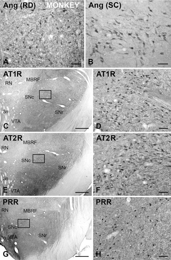
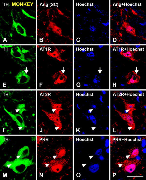
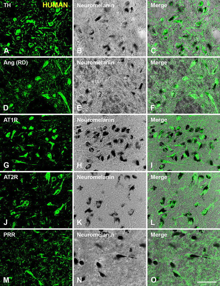
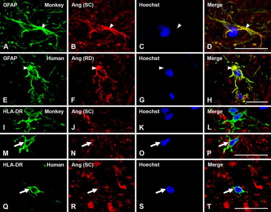
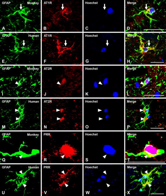
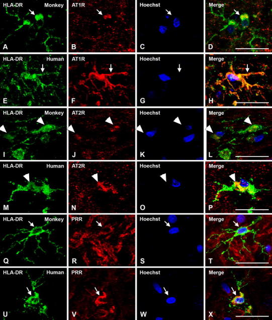
We have previously obtained in rodents a considerable amount of data suggesting a major role for the brain renin-angiotensin system (RAS) in dopaminergic neuron degeneration and potentially in Parkinson's disease. However, the presence of a local RAS has not been demonstrated in the monkey or the human substantia nigra compacta (SNc). The present study demonstrates the presence of major RAS components in dopaminergic neurons, astrocytes and microglia in both the monkey and the human SNc. Angiotensin type 1 and 2 and renin-prorenin receptors were located at the surface of dopaminergic neurons and glial cells, as expected for a tissular RAS. However, angiotensinogen and receptors for angiotensin and renin-prorenin were also observed at the cytoplasm and nuclear level, which suggests the presence of an intracrine or intracellular RAS in monkey and human SNc. Although astrocytes and microglia were labeled for angiotensin and prorenin receptors in the normal SNc, most glial cells appeared less immunoreactive than the dopaminergic neurons. However, our previous studies in rodent models of PD and studies in other animal models of brain diseases suggest that the RAS activity is significantly upregulated in glial cells in pathological conditions. The present results together with our previous findings in rodents suggest a major role for the nigral RAS in the normal functioning of the dopaminergic neurons, and in the progression of the dopaminergic degeneration.


Figures

Fig. 1
Fig. 1
Coronal sections through the monkey ventral mesencephalon showing immunoperoxidase labeling for angiotensinogen/angiotensin (a, b), or AT1R (c, d), or AT2R (e, f), or PPR (g, h) show a large number of immunoreactive cells in the substantia nigra compacta. a, b, d, f, h show high magnification photographs of the areas boxed in c, e, g. 3n third cranial nerve, Ang angiotensinogen/angiotensin, AT1R angiotensin II type 1 receptor, AT2R angiotensin II type 2 receptor, MBRF midbrain reticular formation, PRR prorenin/renin receptor, RD R&D systems, RN red nucleus, SC Santa Cruz Biotechnology, SNc substantia nigra pars compacta, SNr substantia nigra pars reticulata, VTA ventral tegmental area. Scale bars 100 µm (a, b, d, f, h); 1 mm (c, e, g)
Fig. 2
Fig. 2
Coronal sections through the human ventral mesencephalon showing immunoperoxidase labeling for angiotensinogen/angiotensin (a, b), or AT1R (c, d), or AT2R (e, f), or PPR (g, h) show a large number of immunoreactive cells in the substantia nigra compacta. a, b, d, f, h show high magnification photographs of the areas boxed in c, e, g. Enlargements of the areas boxed in d, f, h show the presence of neuromelanin granules (arrowheads) together with AT1R, AT2R or PPR immunolabeling (arrows). 3n third cranial nerve, Ang angiotensinogen/angiotensin, AT1R angiotensin II type 1 receptor, AT2R angiotensin II type 2 receptor, PRR prorenin/renin receptor, RD R&D systems, RN red nucleus, SC Santa Cruz Biotechnology, SNc substantia nigra pars compacta, SNr substantia nigra pars reticulata. Scale bars 100 µm (a, b, d, f, h); 1 mm (c, e, g); 50 µm (enlargements in d, f, h)
Fig. 3
Fig. 3
Double immunofluorescence for tyrosine hydroxilase (green dopaminergic neurons) and angiotensinogen/angiotensin (Ang; SC, Santa Cruz Biotechnology) or AT1R or AT2R or PRR (red) in the monkey substantia nigra compacta. Ang (ac) and AT1R (df), AT2R (gi) and PRR (jl) show co-localization (yellow) with the dopaminergic marker TH. Labeling is apparently located at cell surface (arrows in df), neuron nucleus, and cytoplasmic (arrowheads in gl) levels. The cytoplasmic labeling for PRR was weaker than for AT1R and AT2R, but clearly visible at high magnification (arrowheads in jl). Ang angiotensinogen/angiotensin, AT1R angiotensin II type 1 receptor, AT2R angiotensin II type 2 receptor, PRR prorenin/renin receptor, SC Santa Cruz Biotechnology, RD R&D Systems, TH tyrosine hydroxylase Scale bar 100 µm
Fig. 4
Fig. 4
Triple immunofluorescent labeling showing dopaminergic neurons at high magnification in the monkey substantia nigra compacta (TH-ir; a, e, i, m; green), and double-labeled for the nuclear marker Hoechst 33342 (blue) and (red) angiotensinogen/angiotensin (ad), AT1R (eh), AT2R (il) and PRR (mp). Labeling is apparently located at cell surface (arrows in eh), cytoplasmic (arrowheads in ip) and nuclear levels. AT1R labeling is most intense at the cell surface and in the nucleus (eh), AT2R labeling in the cytoplasm (il) and PRR labeling at the nuclear level (mp). The weak cytoplasmic labeling for PRR is shown by arrowheads in mp. Ang angiotensinogen/angiotensin, AT1R angiotensin II type 1 receptor, AT2R angiotensin II type 2 receptor, PRR prorenin/renin receptor, SC Santa Cruz Biotechnology, TH tyrosine hydroxylase. Scale bar 25 µm
Fig. 5
Fig. 5
Immunofluorescent labeling of dopaminergic neurons in the human substantia nigra compacta were identified either with the dopaminergic marker tyrosine hydroxylase or clearly visible neuromelanin granules, which co-localized in dopaminergic neurons as shown in ac. Angiotensinogen/angiotensin (df), AT1R (gi), AT2R (jl), and PRR (mo) were located in all dopaminergic neurons containing neuromelanin granules. Ang angiotensinogen/angiotensin, AT1R angiotensin II type 1 receptor, AT2R angiotensin II type 2 receptor, PRR prorenin/renin receptor, RD R&D Systems, TH tyrosine hydroxylase. Scale bar 100 µm
Fig. 6
Fig. 6
Triple immunofluorescent labeling for the astroglial marker GFAP (ah; green) or the microglial marker HLA-DR (it; green), the nuclear marker Hoechst 33342 (blue), and angiotensinogen/angiotensin (Ang; red) in monkey and human substantia nigra compacta. In astrocytes, labeling is mainly located at the cytoplasmic level. In microglial cells of normal substantia nigra, labeling for angiotensinogen/angiotensin was weak and particularly located at the cell surface (arrows). Ang angiotensinogen/angiotensin, GFAP glial fibrillary acidic protein, HLA-DR human leukocyte antigen DR, SC Santa Cruz Biotechnology, RD R&D Systems. Scale bar 20 µm
Fig. 7
Fig. 7
Triple immunofluorescent labeling for the astroglial marker GFAP (green), the nuclear marker Hoechst 33342 (blue), and AT1R, AT2R or PRR (red) in monkey and human substantia nigra compacta. Labeling for AT1R appeared mostly located at the cell surface (arrows in ah), and AT2R and PRR labeling at the cytoplasm and nuclear levels (arrowheads in ix). AT1R angiotensin II type 1 receptor, AT2R angiotensin II type 2 receptor, GFAP glial fibrillary acidic protein, PRR prorenin/renin receptor. Scale bar 20 µm
Fig. 8
Fig. 8
Triple immunofluorescent labeling for the microglial marker HLA-DR (green), the nuclear marker Hoechst 33342 (blue), and AT1R, AT2R or PRR receptors (red) in monkey and human substantia nigra compacta. Labeling for AT1R is apparent at the cell surface, cytoplasm and nuclear levels (ah, arrows); labeling for AT2R is apparent mainly at the cytoplasmic level (ip, arrowheads), and PRR mainly at the cell surface level (qx, arrows). AT1R angiotensin II type 1 receptor, AT2R angiotensin II type 2 receptor, HLA-DR human leukocyte antigen DR, PRR prorenin/renin receptor. Scale bar 20 µm

References

    1. Allen AM, MacGregor DP, Chai SY, Donnan GA, Kaczymarzyk S, Richardson K, Kalnins R, Ireton J, Mendelsohn FAO. Angiotensin II receptor binding associated with nigrostriatal dopaminergic neurons in human basal ganglia. Ann Neurol. 1992;32:339–344. doi: 10.1002/ana.410320306. - DOI - PubMed
    1. Allen AM, Moeller I, Jenkins A, Zhuo J, Aldred GP, Chai SY, Mendelsohn FAO. Angiotensin receptors in the nervous system. Brain Res Bull. 1998;47:17–18. doi: 10.1016/S0361-9230(98)00039-2. - DOI - PubMed
    1. Baker KM, Chernin MI, Schreiber T, Sanghi S, Haiderzaidi S, Booz GW, Dostal DE, Kumar R. Evidence of a novel intracrine mechanism in angiotensin II-induced cardiac hypertrophy. Regul Pept. 2004;120:5–13. doi: 10.1016/j.regpep.2004.04.004. - DOI - PubMed
    1. Benigni A, Corna D, Zoja C, Sonzogni A, Latini R, Salio M, Conti S, Rottoli D, Longaretti L, Cassis P, Morigi M, Coffman TM, Remuzzi G. Disruption of the Ang II type 1 receptor promotes longevity in mice. J Clin Invest. 2009;119:524–530. doi: 10.1172/JCI36703. - DOI - PMC - PubMed
    1. Chabrashvili T, Kitiyakara C, Blau J, Karber A, Islam S, Welch WJ, Wilcox CF. Effect of Ang II type 1 and 2 receptors on oxidative stress, renal NAD(P)H oxidase, and SOD expression. Am J Physiol Regu Integr Comp Physiol. 2003;285:R117–R124. - PubMed

Publication types

MeSH terms

LinkOut - more resources