. 2012 Jun;27(6):1357-67.
doi: 10.1002/jbmr.1588.

The gut microbiota regulates bone mass in mice

Affiliations
Free PMC article

The gut microbiota regulates bone mass in mice

Klara Sjögren et al. J Bone Miner Res. 2012 Jun.
Free PMC article

Abstract

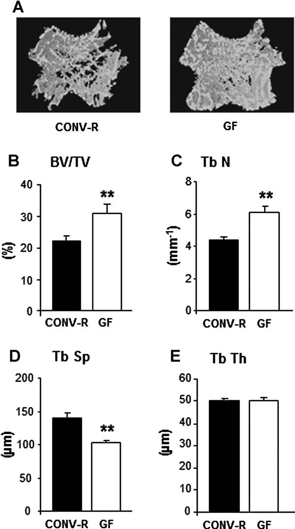
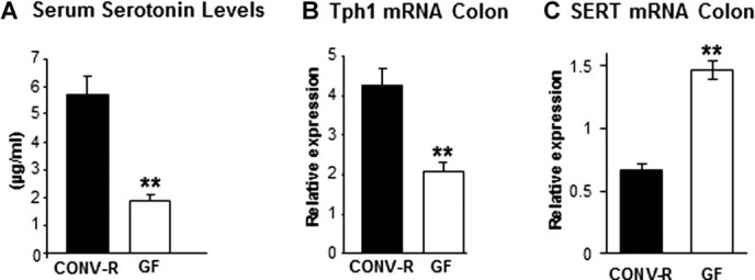
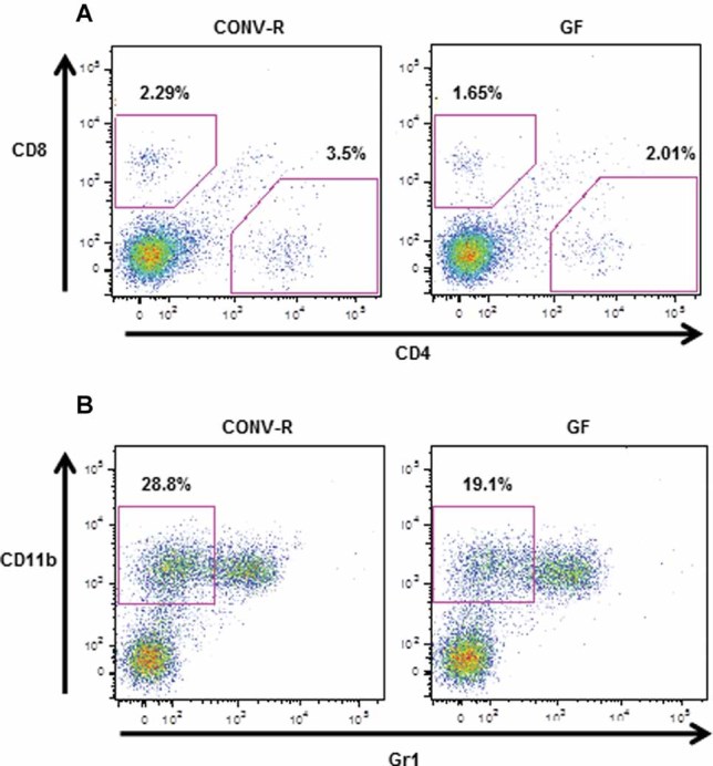
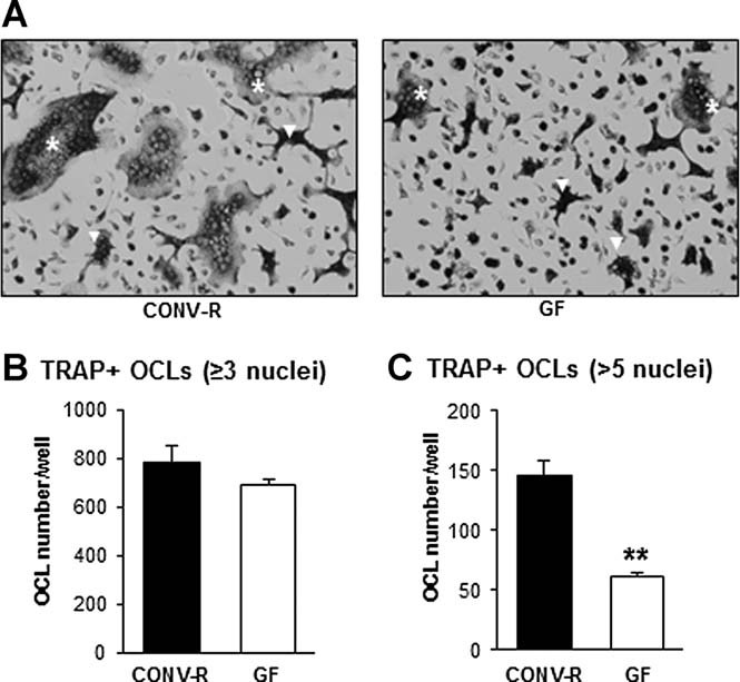
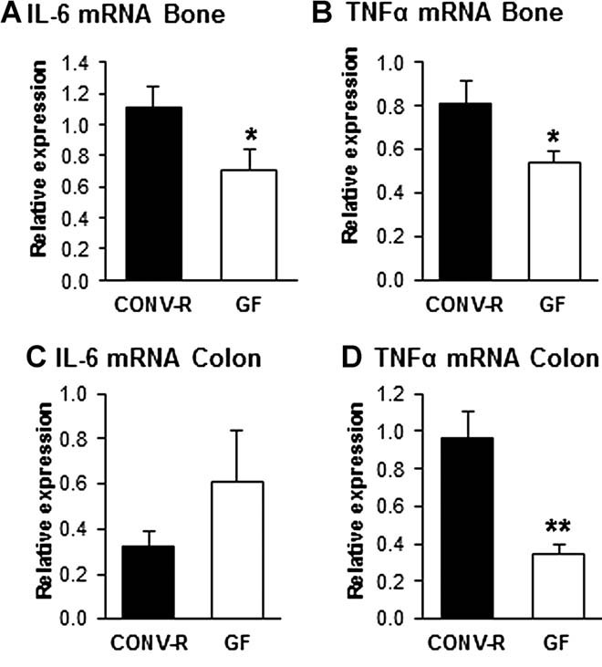
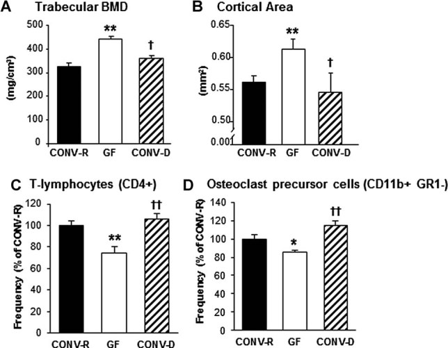
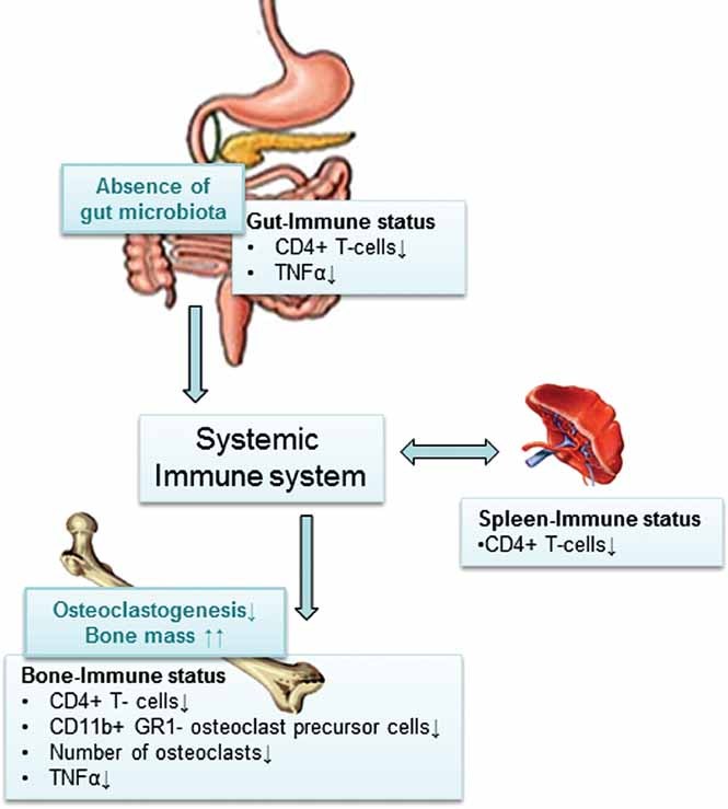
The gut microbiota modulates host metabolism and development of immune status. Here we show that the gut microbiota is also a major regulator of bone mass in mice. Germ-free (GF) mice exhibit increased bone mass associated with reduced number of osteoclasts per bone surface compared with conventionally raised (CONV-R) mice. Colonization of GF mice with a normal gut microbiota normalizes bone mass. Furthermore, GF mice have decreased frequency of CD4(+) T cells and CD11b(+) /GR 1 osteoclast precursor cells in bone marrow, which could be normalized by colonization. GF mice exhibited reduced expression of inflammatory cytokines in bone and bone marrow compared with CONV-R mice. In summary, the gut microbiota regulates bone mass in mice, and we provide evidence for a mechanism involving altered immune status in bone and thereby affected osteoclast-mediated bone resorption. Further studies are required to evaluate the gut microbiota as a novel therapeutic target for osteoporosis.


Figures

Fig. 1
Fig. 1
Absence of gut microbiota leads to increased bone mass in mice. Trabecular bone parameters were analyzed by µCT in the distal metaphyseal region of femur from 7-week-old germ-free (GF) and conventionally raised (CONV-R) female mice. (A) Representative µCT images of one trabecular section from each group. (B) BV/TV (%), trabecular bone volume as a percentage of tissue volume. (C) Tb.N (mm−1), trabecular number. (D) Tb.Sp (µm), trabecular separation. (E) Tb.Th (µm), trabecular thickness. Values are given as mean ± SEM, n = 8 to 14. *p ≤ 0.05; **p ≤ 0.01 versus CONV-R, Student's t test.
Fig. 2
Fig. 2
GF mice have decreased peripheral serotonin synthesis. (A) Serum serotonin levels in 7-week-old GF and CONV-R female mice. (B) qRT-PCR analysis of the expression of tryptophan hydroxylase-1 (Tph1) in proximal colon of 9-week-old GF and CONV-R female mice. (C) qRT-PCR analysis of the expression of the serotonin transporter (SERT) in proximal colon of 9-week-old GF and CONV-R female mice. Values are given as mean ± SEM, n = 5 to 14. **p ≤ 0.01 versus CONV-R, Student's t test.
Fig. 3
Fig. 3
GF mice have a decreased frequency of CD4 T cells and osteoclast (OCL) precursor cells (CD11b+/Gr1) in bone marrow. Femur bone marrow cells from 7-week-old GF and CONV-R female mice were stained with antibodies recognizing CD8, CD4, CD11b, and Gr1 and analyzed by flow cytometry, gating as shown. (A) Values represent the percentage of CD8+ and CD4+ cells in the total bone marrow population. Data are representative of two independent experiments. (B) Values represent the percentage of CD11b+/Gr1 cells (OCL precursor cells; see text for details) in the total bone marrow population. Data are representative of two independent experiments.
Fig. 4
Fig. 4
Osteoclast formation in vitro is decreased in bone marrow cultures from GF mice. Bone marrow cells were flushed from femur and tibias from 8-week-old GF and CONV-R female mice, cultured for 4 days, and then stained for tartrate-resistant acid phosphatase (TRAP). (A) Representative images showing TRAP + OCLs in culture from each group. White stars (*) indicate examples of spread OCLs with more than five nuclei, and white arrows (Δ) indicate examples of OCLs with more than three but less than five nuclei. (B) Quantitative data for TRAP-positive cells containing three or more nuclei. (C) Quantitative data for spread TRAP-positive cells containing more than five nuclei. Values are given as mean ± SEM, n = 4 to 5. **p < 0.01 versus CONV-R, Student's t test.
Fig. 5
Fig. 5
GF mice have decreased expression of IL-6 and TNFα in bone. QRT-PCR analysis of the expression of (A) interleukin-6 (IL-6) and (B) tumor necrosis factor alpha (TNFα) in tibias from 7-week-old GF and CONV-R female mice and (C) IL-6 and (D) TNFα in proximal colon of 9-week-old GF and CONV-R female mice. Values are given as mean ± SEM, n = 5 to 14. *p ≤ 0.05 versus CONV-R, Student's t test.
Fig. 6
Fig. 6
Colonization of GF mice with a normal gut microbiota normalizes bone mass and immune status in bone marrow. Analysis of bone and bone marrow in female 7-week-old GF and CONV-R mice and an extra control group consisting of mice that were born GF and then colonized with normal gut microbiota at 3 weeks of age (conventionalized; CONV-D). (A) Trabecular bone mineral density (BMD) measured by pQCT in the distal femur. (B) Cortical cross-sectional bone area measured by µCT in the diaphyseal region of femur. (C) Quantitative data for the frequency of T cells (CD4+) in the total bone marrow population; (D) Quantitative data for the frequency of CD11b+/Gr1 cells (OCL precursor cells; see text for details) in the total bone marrow population. Values are given as mean ± SEM, n = 4 to 14 in each group. **p ≤ 0.01, GF versus CONV-R; †p ≤ 0.05, ††p ≤ 0.01, CONV-D versus GF, ANOVA followed by Tukey's post hoc test.
Fig. 7
Fig. 7
Proposed mechanism for how the gut microbiota regulates bone mass. Absence of gut microbiota leads to increased bone mass associated with reduced number of OCLs per bone surface, decreased frequency of CD4+ T cells and OCL precursor cells in bone marrow, and reduced expression of the osteolytic cytokine TNFα in bone. Earlier studies have shown that GF animals have immature mucosal immune systems with hypoplastic Peyer's patches containing few germinal centers and reduced number of IgA-producing plasma cells and lamina propria CD4+ T cells. Furthermore, GF mice have reduced number of CD4+ T cells in the spleen and fewer and smaller germinal centers within the spleen, suggesting that the gut microbiota is capable of shaping systemic immunity., We propose that the increased bone mass is caused by fewer CD4+ cells recirculating in blood and secondary lymphoid tissue, resulting in a decreased frequency of CD4+ T cells in bone marrow associated with a decreased expression of inflammatory cytokines and less osteoclastogenesis in the absence of gut microbiota.

Comment in

References

    1. Qin J, Li R, Raes J, Arumugam M, Burgdorf KS, Manichanh C, Nielsen T, Pons N, Levenez F, Yamada T, Mende DR, Li J, Xu J, Li S, Li D, Cao J, Wang B, Liang H, Zheng H, Xie Y, Tap J, Lepage P, Bertalan M, Batto JM, Hansen T, Le Paslier D, Linneberg A, Nielsen HB, Pelletier E, Renault P, Sicheritz-Ponten T, Turner K, Zhu H, Yu C, Jian M, Zhou Y, Li Y, Zhang X, Qin N, Yang H, Wang J, Brunak S, Dore J, Guarner F, Kristiansen K, Pedersen O, Parkhill J, Weissenbach J, Bork P, Ehrlich SD. A human gut microbial gene catalogue established by metagenomic sequencing. Nature. 2010;464(7285):59–65. - PMC - PubMed
    1. Ley RE, Hamady M, Lozupone C, Turnbaugh PJ, Ramey RR, Bircher JS, Schlegel ML, Tucker TA, Schrenzel MD, Knight R, Gordon JI. Evolution of mammals and their gut microbes. Science. 2008;320(5883):1647–51. - PMC - PubMed
    1. Rhee SH, Pothoulakis C, Mayer EA. Principles and clinical implications of the brain-gut-enteric microbiota axis. Nat Rev Gastroenterol Hepatol. 2009;6(5):306–14. - PMC - PubMed
    1. Backhed F, Ding H, Wang T, Hooper LV, Koh GY, Nagy A, Semenkovich CF, Gordon JI. The gut microbiota as an environmental factor that regulates fat storage. Proc Natl Acad Sci USA. 2004;101(44):15718–23. - PMC - PubMed
    1. Jarchum I, Pamer EG. Regulation of innate and adaptive immunity by the commensal microbiota. Curr Opin Immunol. 2011;23(3):353–60. - PMC - PubMed

Publication types

MeSH terms