. 2012 May 3;119(18):4142-51.
doi: 10.1182/blood-2011-09-381970. Epub 2012 Mar 9.

The FA pathway counteracts oxidative stress through selective protection of antioxidant defense gene promoters

Affiliations

The FA pathway counteracts oxidative stress through selective protection of antioxidant defense gene promoters

Wei Du et al. Blood. .

Abstract

Oxidative stress has been implicated in the pathogenesis of many human diseases including Fanconi anemia (FA), a genetic disorder associated with BM failure and cancer. Here we show that major antioxidant defense genes are down-regulated in FA patients, and that gene down-regulation is selectively associated with increased oxidative DNA damage in the promoters of the antioxidant defense genes. Assessment of promoter activity and DNA damage repair kinetics shows that increased initial damage, rather than a reduced repair rate, contributes to the augmented oxidative DNA damage. Mechanistically, FA proteins act in concert with the chromatin-remodeling factor BRG1 to protect the promoters of antioxidant defense genes from oxidative damage. Specifically, BRG1 binds to the promoters of the antioxidant defense genes at steady state. On challenge with oxidative stress, FA proteins are recruited to promoter DNA, which correlates with significant increase in the binding of BRG1 within promoter regions. In addition, oxidative stress-induced FANCD2 ubiquitination is required for the formation of a FA-BRG1-promoter complex. Taken together, these data identify a role for the FA pathway in cellular antioxidant defense.


Figures
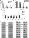
Figure 1
Figure 1
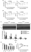
Down-regulation of antioxidant genes in FA BM cells. (A) Hypersensitivity of FA BM progenitors to H2O2. BM cells from 5 FA-A and 2 FA-C patients, as well as 5 healthy donors, were treated with 100μM H2O2 for 2 hours followed by a colony-forming assay. Colony numbers were counted 10 days after plating. Results are means ± SD of 3 independent experiments. (B) Hypersensitivity of FA LCLs to H2O2. FA-A LCLs and cells corrected with the FANCA gene were treated with increasing doses of H2O2 for 2 hours followed by culture in fresh medium for an additional 24 hours. Cells were then analyzed for cell survival. (C) BM cells from 5 FA-A, 1 FA-B, 2 FA-C, 1 FA-D1, 1 FA-I, and 1 FA-J patients and healthy donors were used for pathway-specific (oxidative stress and antioxidant defense) array analysis. Eighty-four genes involved in antioxidative stress response were analyzed. The data represent the fold down-regulation of the indicated genes in FA samples relative to healthy donors. Fold down-regulation = −1/(fold difference), where fold difference = [2−ΔΔCt (FA)]/[2−ΔΔCt (healthy)]. (D) RT-PCR analysis of antioxidant gene expression in FA-A BM cells. RNA was extracted from BM cells from 2 FA-A patients and 2 healthy donors followed by RT-PCR analysis. (E) Western analysis of antioxidant gene products in FA-A BM cells. Protein lysates were prepared from cells described in panel B followed by Western blot analysis using the indicated Abs. Relative mRNA and protein levels were quantified using ImageJ software (NIH). The results were plotted after normalization with β-actin as an internal control.
Figure 2
Figure 2
Down-regulation of antioxidant genes is associated with a selective increase in promoter DNA damage in FA cells. (A) Increased promoter DNA damage in antioxidant genes in FA-A cells. FA-A cells transduced with empty vector or cDNA-encoding FANCA, as well as a normal control, were treated with 100μM H2O2 for 2 hours followed by 12 hours of culture in fresh medium. Genomic DNA was isolated followed by FPG cleavage and qPCR using primers specific for the promoters of the indicated genes. The percentage of intact DNA represents the ratio of PCR products after Fpg cleavage to those present in uncleaved DNA. (B) DNA damage in the coding sequences of antioxidant defense genes. The same analysis was applied as described in panel A using primers specific for exons of the indicated genes. (C-D) Increased 8-oxodG accumulation in the promoters of antioxidant genes in FA cells. FA-A or gene-corrected cells were treated with increasing doses of H2O2 for 2 hours and released into fresh medium for another 12 hours, followed by ChIP using an Ab against 8-oxodG. Precipitated samples were then subjected to PCR using primers specific for promoter regions of (C) GPX1 or (D) TXNRD1 gene. Representative images (left) and quantifications (right) are shown. The intensities of DNA bands were quantified using ImageJ software (NIH). Results are means ± SD of 3 independent experiments.
Figure 3
Figure 3
Increased initial oxidative DNA damage to the promoters of antioxidant genes in FA cells. (A) Repair kinetics of oxidative damage to GPX1 promoter. FA-A cells or gene-corrected cells were treated with H2O2 for 2 hours and released for the indicated time intervals followed by genomic DNA or RNA isolation. Samples were then subjected to (left) DNA-damage assay or (right) RT-PCR. (B) Repair kinetics of oxidative damage to GSTP1 promoter. Samples described in panel A were then subjected to (left) DNA-damage assay or (right) RT-PCR. Percentage of intact DNA is the ratio of PCR products after Fpg cleavage to those present in uncleaved DNA. (C) Increased initial oxidative DNA damage in FA-A cells. Cells described in panel A were used for ChIP using an Ab against 8-oxodG and PCR using primers specific for (left) GPX1 or (right) GSTP1 promoter. Representative images (top) and quantifications (bottom) are shown. Results are means ± SD of 3 independent experiments. (D) Repair efficiency as determined by host cell-reactivation assay. The pSSG-promoter reporter vector containing promoter regions of antioxidant gene GCLC, GPX1, GSTP1, or TXNRD1, as well as control gene GAPDH or β-tubulin, were treated with 100μM H2O2 for 1 hour in vitro and then transfected into normal, FA-A, or gene-corrected fibroblasts followed by determination of luciferase activity. Results are means ± SD of 3 independent experiments. (E) Repair kinetics of oxidative damage to naked promoter DNA. Genomic DNA was isolated from cells described in panel D followed by Fpg cleavage and qPCR using primers specific for the cloned GPX1 promoter. The level of intact DNA represents the efficiency of repair.
Figure 4
Figure 4
Oxidative stress-induced formation of a FA-BRG1–promoter complex. (A) FANCD2 does not bind to the promoters of antioxidant genes in unstressed cells. Untreated normal lymphoblasts were subjected to a ChIP assay using Abs against FANCD2 or BRG1. PCR amplification was performed using primers specific for the promoters of indicated antioxidant or housekeeping genes. (B) FANCD2 is recruited to the GPX1and TXNRD1 promoter regions after H2O2 treatment. Normal cells were treated with 100μM H2O2 for 2 hours then released for the indicated time intervals. Proteins were extracted at different time points, followed by a ChIP assay using Abs against FANCD2 or BRG1. Precipitated samples were then subjected to PCR using primers for the promoters of GPX1 or TXNRD1. Representative images (top) and quantifications (botton) were shown. The intensity of the DNA bands was quantified using ImageJ software (NIH). Results are means ± SD of 3 independent experiments. (C) FA-BRG1-promoter complex was absent in FA cells. FA-A cells were treated with 100μM H2O2 for 2 hours then released for the indicated time intervals. Proteins were extracted at different time points, followed by a ChIP assay using Abs against FANCD2 or BRG1. Precipitated samples were then subjected to PCR using primers for the promoters of GPX1 or TXNRD1. (D) Oxidative stress induces accumulation of acetylated histone in the promoters of antioxidant genes of both normal and FA cells. Normal and FA-A cells were treated with or without 100μM H2O2 for 2 hours followed by release into fresh medium. Cells were then subjected to ChIP assay using Abs specific for acetylated histone H3K9/14 (Ac-H3K9/14) or methylated histone H3K9 (Me-H3K9) followed by PCR using primers for the promoter regions of GPX1, TXNRD1, or β-tubulin. Representative images (top) and quantifications (bottom) are shown. The intensity of the DNA bands was quantified using ImageJ software (NIH). Results are means ± SD of 3 independent experiments. (E) Repair kinetics of oxidative damage in BRG1-bound antioxidant gene promoter. FA-A and gene-corrected cells were treated with or without H2O2 for 2 hours and released into fresh medium for up to 24 hours. ChIP assay using Abs against BRG1 was performed, and the bound DNA fragments were subjected to the Fpg cleavage/PCR-based DNA repair assay using primers specific for the promoter of (left) GPX1 or (right) TXNRD1. Percentage of intact DNA represents the ratio of PCR products after Fpg cleavage to those present in uncleaved DNA.
Figure 5
Figure 5
Binding of FANCD2 to promoters is independent of BRG1. (A) Knockdown of BRG1. Normal lymphoblasts expressing a shRNA for Brg1 or a control shRNA were treated with or without 100μM H2O2 for 2 hours. Cell extracts were then subjected to Western blotting using Abs against BRG1 and actin. (B) Binding of FANCD2 to promoters is independent of BRG1. Cells described in panel A were treated with or without H2O2, followed by a ChIP assay using Abs against FANCD2. PCR was performed using primers specific for the promoter regions of GPX1, TXNRD1, GAPDH, or β-tubulin.
Figure 6
Figure 6
FANCD2 ubiquitination is required for the formation of FA-BRG1–promoter complex. (A) Reconstitution of the FA-D2 cells with WT FANCD2 or the nonubiquitinated FANCD2-K561R mutant. FANCD2-deficient PD20 cell were transduced with retrovirus-expressing empty vector, WT-FANCD2, or the FANCD2-K561R mutant followed by puromycin selection. Stable cell lines were treated with or without H2O2 followed by Western analysis using Abs against FANCD2, FANCA, BRG1, or β-actin. (B) Oxidative stress induces chromatin loading of BRG1 and FA proteins in FANCD2-corrected cells. Cells described in panel A were treated with or without H2O2 followed by chromatin fractionation. Chromatin extracts were then analyzed by Western blotting with Abs against BRG1, FANCA, FANCC, FANCD2, FANCG, or FOXO3a. Histone H2A was included as a loading control. (C) FANCD2 ubiquitination is required for the formation of the FA-BRG1-DNA complex. Cells described in panel A were transduced with retrovirus expressing Flag-tagged FANCA, followed by cell sorting for GFP. Sorted cells were then treated with or without H2O2 for 2 hours and released for the indicated hours. ChIP assays using Abs against Flag or BRG1 were followed by PCR amplification using primers specific for the promoter of GPX1. (D) FANCD2 ubiquitination is required for the protection of antioxidant gene promoter DNA from oxidative damage. Cells described in panel C were treated with or without H2O2 for 2 hours and released into fresh medium for an additional 12 hours. ChIP assay using Ab against BRG1 was performed, and bound DNA fragments were subjected to the Fpg cleavage/PCR-based DNA-repair assay using primers specific for the promoter of GPX1. The percentage of intact DNA represents the ratio of PCR products after Fpg cleavage to those present in uncleaved DNA.

References

    1. Klaunig JE, Kamendulis LM, Hocevar BA. Oxidative stress and oxidative damage in carcinogenesis. Toxicol Pathol. 2010;38(1):96–109. - PubMed
    1. Weiss JM, Goode EL, Ladiges WC, Ulrich CM. Polymorphic variation in hOGG1 and risk of cancer: A review of the functional and epidemiologic literature. Mol Carcinog. 2005;42(3):127–141. - PubMed
    1. Tanaka H, Fujita N, Sugimoto R, et al. Hepatic oxidative DNA damage is associated with increased risk for hepatocellular carcinoma in chronic hepatitis C. Br J Cancer. 2008;98(3):580–586. - PMC - PubMed
    1. Gottschling BC, Maronpot RR, Hailey JR, et al. The role of oxidative stress in indium phosphide-induced lung carcinogenesis in rats. Toxicol Sci. 2001;64(1):28–40. - PubMed
    1. Muguruma M, Unami A, Kanki M, et al. Possible involvement of oxidative stress in piperonyl butoxide induced hepatocarcinogenesis in rats. Toxicology. 2007;236(1-2):61–75. - PubMed

Publication types

MeSH terms