. 2012 May 17;119(20):4786-97.
doi: 10.1182/blood-2011-10-387522. Epub 2012 Mar 9.

Regulation of acute graft-versus-host disease by microRNA-155

Affiliations

Regulation of acute graft-versus-host disease by microRNA-155

Parvathi Ranganathan et al. Blood. .

Abstract

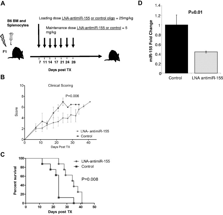
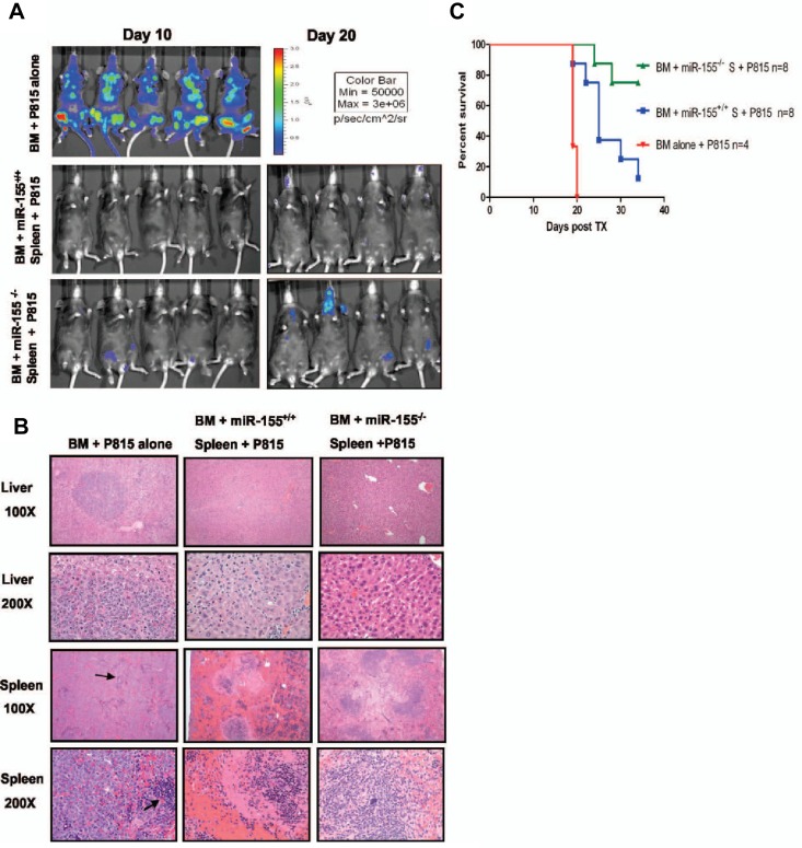
Acute graft-versus-host disease (aGVHD) remains a major complication of allogeneic hematopoietic stem cell transplant (alloHSCT), underscoring the need to further elucidate its mechanisms and develop novel treatments. Based on recent observations that microRNA-155 (miR-155) is up-regulated during T-cell activation, we hypothesized that miR-155 is involved in the modulation of aGVHD. Here we show that miR-155 expression was up-regulated in T cells from mice developing aGVHD after alloHSCT. Mice receiving miR-155-deficient donor lymphocytes had markedly reduced lethal aGVHD, whereas lethal aGVHD developed rapidly in mice recipients of miR-155 overexpressing T cells. Blocking miR-155 expression using a synthetic anti-miR-155 after alloHSCT decreased aGVHD severity and prolonged survival in mice. Finally, miR-155 up-regulation was shown in specimens from patients with pathologic evidence of intestinal aGVHD. Altogether, our data indicate a role for miR-155 in the regulation of GVHD and point to miR-155 as a novel target for therapeutic intervention in this disease.


Figures

Figure 1
Figure 1
miR-155 expression is up-regulated in effectors T cells of mice recipients with aGVHD. (A) Schematic presentation showing aGVHD murine model used. (B) Histopathologic evaluation of a representative liver sample collected from a mouse with clinical score of GVHD of more than or equal to 7. This section was stained with hematoxylin and eosin (original magnification ×40 and ×400). (C) Average miR-155 expression in CD4+ CD62L T cells isolated from the spleen of 3 recipient mice with aGVHD (BM + spleen) or controls (BM alone). The results shown represent the average of 3 independent mouse samples performed each in triplicate. Values are expressed as relative miRNA expression after 2ΔCt calculations and normalization with sno135. Bars represent SD. (D) Hierarchical clustering analysis of miRNA expression obtained using Nanostring from CD4+ CD62L T cells isolated from the spleen of recipient mice with aGVHD (BM + spleen) or controls (BM alone). Color areas indicate relative expression of each miRNA among the 2 type of samples (red high, blue low expression). (E) Histopathologic and molecular evaluation of a representative liver sample collected from a mouse with clinical score of GVHD of more than or equal to 7 or a control mouse (BM alone). At least 3 mice in each group were evaluated for staining with LNA anti–miR-155 and scramble control. These sections were stained with LNA anti–miR-155 probes or scrambled controls. Dark staining indicates positivity (original magnification ×400). Note the strong staining of the mononuclear cells in the periportal lymphoid aggregate in the BM + spleen mouse that is lost in the same cells in the serial section with the scrambled probe.
Figure 2
Figure 2
Recipients from miR-155–deficient splenocytes do not develop severe aGVHD and have increased survival. (A) Schematic presentation showing the different donor cells used in lethally irradiated F1 mice. (B) Clinical scores from the different recipient mice groups after transplantation. (C) Survival rate of lethally irradiated mice receiving BM alone, BM + miR-155−/− spleen, BM + miR-155+/+ spleen, or XRT alone. Data represent 2 independent experiments. (D) Histopathologic GVHD scores of recipient tissues. A second cohort of mice was used for histopathologic analysis. Fifteen days after transplantation (except for XRT group), mice from the indicated groups were killed and sections of the large bowel and liver were stained with H&E. Data are pooled from 2 independent experiments, representing approximately 6 or 7 animals per group, the XRT group that had 4 mice. There was no significant difference in the liver GVHD scores between the BM alone and BM + miR-155−/− spleen recipients (P = .07). The GVHD scores in the gut were higher in the BM + miR-155−/− spleen recipients versus the BM alone (P = .04). (E) Histopathologic evaluation of representative liver and large-bowel samples collected from the different recipient mice groups at day 15 after transplantation. The arrows in the large-bowel slide from the B6 BM + miR-155+/+ spleen F1 recipients showed lymphoplasmacytic infiltration in the lamina propia with frequent apoptosis The arrows in the liver slide from the same F1 recipients showed abundant lymphocytes infiltrating around the portal vein. No significant pathology is shown in BM alone and miR-155−/− groups. (F) Clinical GVHD scores after transplantation from lethally irradiated B10.BR recipients transplanted with BM alone, BM + miR-155−/−, or BM + miR-155+/+ spleen. (G) Survival rate of the different recipient B10.BR mice groups. (H) TNF-α levels measured in the serum of lethally irradiated F1 recipients of BM alone, BM + miR-155−/− spleen, and BM + miR-155+/+ spleen, 15 days after transplantation. Samples were assayed in triplicate and results are shown as the means from 4 biologic samples within a group.
Figure 3
Figure 3
Recipient mice from donor splenocytes overexpressing miR-155 in T cells exhibit rapidly evolving aGVHD and short survival. (A) Schematic presentation showing the construct used to generate the LCK-miR-155 mice. (B) miR-155 expression (fold change) in the thymus and spleen from age- and sex-matched B6 LCK–miR-155 transgenic mice and B6 miR-155−/− mice. Bars represent SD. (C) Schematic presentation depicting the different recipients F1 mice groups. (D) Clinical GVHD scores after transplantation from lethally irradiated F1 recipients transplanted with BM alone (n = 8), BM + LCK–miR-155 (n = 8), or BM + miR-155+/+ (n = 8) splenocytes. P values were obtained by using t test. (E) Survival rate of the different recipient F1 mice groups. Comparisons between BM + miR-155−/− spleen and BM + miR-155+/+ spleen survival curves were performed using log-rank test. (F) Histopathologic GVHD scores of recipient tissues. A second cohort of mice was used for histopathologic analysis. Fourteen days after transplantation, mice from the indicated groups were killed and sections of the large bowel and liver were stained with H&E. P values were obtained by using t test. (G) Histopathologic evaluation of representative liver samples obtained from recipients of BM alone, BM + miR-155+/+ spleen, and BM + LCK-miR-155 donor cells. Slides were stained with H&E. The arrows indicate lymphocytic infiltration in the recipient livers characteristic of GVHD.
Figure 4
Figure 4
miR-155 modulates chemokine receptor expression and migration of allogeneic donor T cells. On days 4 and 21 after BMT, splenocytes were harvested from F1 recipients and H2Kd- CD4+/CD8+ donor cells were analyzed by flow cytometry for chemokine receptor expression. Chemokine receptor expression was quantified by measuring mean fluorescence intensity (MFI). P values < .05 were considered significant. n = at least 5 mice per group at each time point. (A) Expression of CXCR4 on CD4+ and CD8+ WT and miR-155−/− donor T cells on day 21 after transplantation. (B) Expression of S1P1 on CD4+ WT and miR-155−/− donor T cells on day 21 after transplantation. (C) Expression of CCR5 on CD4+ and CD8+ WT and miR-155−/− donor T cells on day 21 after transplantation. (D) Average IL-12Rb1 mRNA expression in untouched T cells isolated from spleens of recipient mice on day 21 after transplantation by RT-PCR. The results shown represent the average of 9 independent mouse samples performed each in triplicate. Values are expressed as relative expression after 2ΔCt calculations and normalization with 18s RNA. Bars represent SD. (E) Average STAT-4 mRNA expression in untouched T cells isolated from spleens of recipient mice on day 21 after transplantation by RT-PCR. (F) Average IFN-γ mRNA expression in untouched T cells isolated from spleens of recipient mice on day 21 after transplantation by RT-PCR. (G) Average CCR5 mRNA expression in untouched T cells isolated from spleens of recipient mice on day 21 after transplantation by RT-PCR.
Figure 5
Figure 5
Treatment with LNA–anti–miR-155 decreases the severity of aGVHD and prolongs the survival of MHC-mismatched recipient mice. (A) Schematic presentation of the schedule of oligonucleotide injections after transplantation. Briefly, lethally irradiated recipient F1 mice transplanted with BM (5 × 10⁶) plus B6 WT spleen cells (20 × 106) were treated with LNA–anti–miR-155 (n = 10) or control LNA oligos (n = 10) starting at day 7 with a loading dose 25 mg/kg followed by 5 mg/kg intravenously twice weekly up to day 30 after infusion of donor B6 splenocytes. (A) Clinical GVHD scores after transplantation from lethally irradiated F1 recipients transplanted with B6 donor BM and splenocytes and treated with LNA–anti–miR-155 or control oligonucleotide. P values were obtained by using t test. (B) Survival rate of the different recipient F1 mice groups. Comparisons between survival curves were performed using log-rank test. (D) miR-155 expression in spleen from LNA–anti–miR-155 or control treated mice as measured by RT-PCR. Data represent the average of 3 mice. Experiments performed in triplicate. Bars represent SE.
Figure 6
Figure 6
miR-155 deficiency in donor T cells does not abrogate GVL effects. (A) Whole body bioluminescent signal intensity of recipient mice. Mice were imaged on day 10 and day 20. (B) Histopathologic analysis of spleen and liver tissues of the different mice cohorts. Histopathologic examination at lower power (original magnification ×100) revealed extensive nodular leukemic infiltration in the liver in the BM alone recipients, whereas no infiltrates were seen in recipients who received splenocytes, either from WT or miR-155–deficient mice (top panels). Middle top panels: The same liver sections are shown at larger magnification (original magnification ×200). Note in the BM alone the many large anaplastic cells with pleomorphic, multilobular large nuclei and frequent mitosis figures. Last 2 bottom panels: Spleen sections at lower (top) and higher (bottom) magnification. Note the complete replacement of the spleen by the large anaplastic cells in the BM alone group. The arrows point to normal spleen. There were no leukemic infiltrations in the recipient groups who received either WT or miR-155–deficient splenocytes. (C) Survival of lethally irradiated mice transplanted with WT or miR-155–deficient splenocytes plus BM P815 leukemic cells. Survival comparisons were performed using the log-rank test.
Figure 7
Figure 7
miR-155 expression is up-regulated in the intestinal tract from patients with aGVHD. Histopathologic assessment of human small- and large-bowel samples from patients with aGVHD or from control patients who had a bowel biopsy, but no pathology was observed (controls). In situ hybridization was performed using a digoxigenin-labeled LNA-modified probe complementary to miR-155 or a scrambled LNA control probe. (A) The colon tissue shows marked inflammation with a loss of glands and concomitant erosion. Note that the many inflammatory cells in the lamina propria were positive for miR-155 (dark staining; original magnification ×200). (B) The miR-155 in situ hybridization signal is lost in the same cells in the serial section on using the LNA scramble control probe. (C) A section of ileum shows a normal villous to the left side of the panel, and the loss of the villi in the rest of the section. Note the strong signal in the inflammatory cells in the area of the damaged villi with the miR-155 probe (original magnification ×200). (D) Negative control for panel C, showing loss of the signal with the scrambled probe in the same cells in the serial section. (E-F) The area to the right of the panel C is magnified (original magnification ×400) in panel E (miR-155) and panel F (scrambled probe) to underscore the localization of the signal to mononuclear cells. (G) An earlier stage of GVHD with degenerated/regenerating glands with mononuclear infiltrates, which are primarily located in the lamina propria adjacent to the damaged glands. The miR-155 signal is rare in the damaged epithelial cells but is strong in the adjacent inflammatory cells as shown in higher magnification (×1000) (H) where mononuclear infiltrate into the epithelia, typical of GVHD, is evident. (I-J) Normal colon mucosa negative for miR-155 staining (original magnification ×400).

Similar articles

Cited by

References

    1. Ferrara JL, Levine JE, Reddy P, Holler E. Graft-versus-host disease. Lancet. 2009;373(9674):1550–1561. - PMC - PubMed
    1. Socie G, Blazar BR. Acute graft-versus-host disease: from the bench to the bedside. Blood. 2009;114(20):4327–4336. - PMC - PubMed
    1. Banerjee A, Schambach F, DeJong CS, Hammond SM, Reiner SL. Micro-RNA-155 inhibits IFN-gamma signaling in CD4+ T cells. Eur J Immunol. 2010;40(1):225–231. - PMC - PubMed
    1. Haasch D, Chen YW, Reilly RM, et al. T-cell activation induces a noncoding RNA transcript sensitive to inhibition by immunosuppressant drugs and encoded by the proto-oncogene, BIC. Cell Immunol. 2002;217(1):78–86. - PubMed
    1. Rodriguez A, Vigorito E, Clare S, et al. Requirement of bic/microRNA-155 for normal immune function. Science. 2007;316(5824):608–611. - PMC - PubMed

Publication types

MeSH terms