Review
. 2012;13(2):1951-2011.
doi: 10.3390/ijms13021951. Epub 2012 Feb 13.

Tumor heterogeneity: mechanisms and bases for a reliable application of molecular marker design

Affiliations
Review

Tumor heterogeneity: mechanisms and bases for a reliable application of molecular marker design

Salvador J Diaz-Cano. Int J Mol Sci. 2012.

Abstract

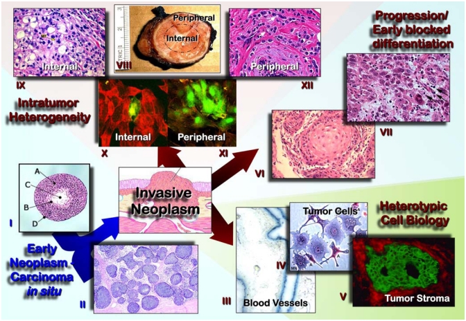
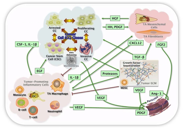
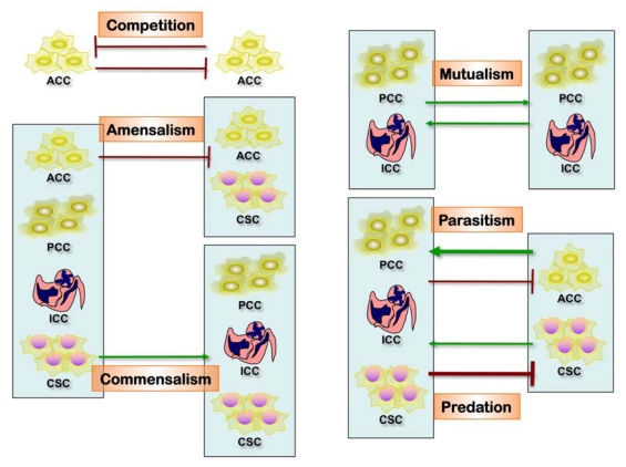
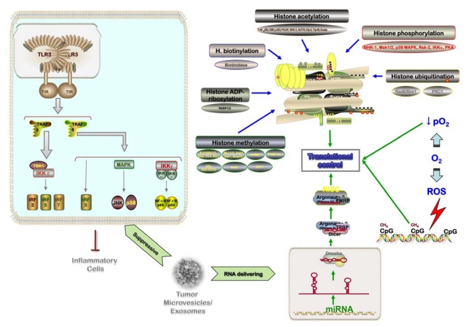
Tumor heterogeneity is a confusing finding in the assessment of neoplasms, potentially resulting in inaccurate diagnostic, prognostic and predictive tests. This tumor heterogeneity is not always a random and unpredictable phenomenon, whose knowledge helps designing better tests. The biologic reasons for this intratumoral heterogeneity would then be important to understand both the natural history of neoplasms and the selection of test samples for reliable analysis. The main factors contributing to intratumoral heterogeneity inducing gene abnormalities or modifying its expression include: the gradient ischemic level within neoplasms, the action of tumor microenvironment (bidirectional interaction between tumor cells and stroma), mechanisms of intercellular transference of genetic information (exosomes), and differential mechanisms of sequence-independent modifications of genetic material and proteins. The intratumoral heterogeneity is at the origin of tumor progression and it is also the byproduct of the selection process during progression. Any analysis of heterogeneity mechanisms must be integrated within the process of segregation of genetic changes in tumor cells during the clonal expansion and progression of neoplasms. The evaluation of these mechanisms must also consider the redundancy and pleiotropism of molecular pathways, for which appropriate surrogate markers would support the presence or not of heterogeneous genetics and the main mechanisms responsible. This knowledge would constitute a solid scientific background for future therapeutic planning.

Keywords: cell segregation; clonal expansion; exosome; metastasis; neoplasm; topographic compartments; tumor heterogeneity; tumor hypoxia; tumor microenvironment; tumor progression.


Figures

Figure 1
Figure 1
Morphological evaluation of neoplasms and development of oncology. Histopathology, as the gold standard of tumor diagnosis, has set up the bases and criteria for the concept of early neoplasms (I showing encapsulated tumor of epithelial cells, A, with central area of edema, C, and a transitional zone, B; D represent the tumor capsule) and carcinomas in situ (II shows a breast lobular carcinoma in situ). Histopathology is contributing to a better understanding of the heterotypic cell biology (III to V reveal tumor cells, blood vessels, inflammatory/immune cells, and stroma; tumor cells and stroma are highlighted by direct immunofluorescence for E-cadherin, green, and integrin, red), the biologic progression/dedifferentiation that has been linked to hypoxic conditions and has been frequently reported in recurrent tumors (VI and VII shows well and poorly differentiated squamous cell carcinoma, respectively, in a recurrent neoplasm), and the intratumor heterogeneity and segregation of tumor cells in predominantly expansile internal compartments IX and X) and predominantly invasive peripheral compartments (XI and XII). The expression of hypoxia up-regulated genes is predominantly observed in the internal compartments (green immunofluorescence for HIF-1α in the example).
Figure 2
Figure 2
Heterotypic cell biology of tumors and microenvironment. Distinct cell types constitute most solid tumors for both tumor parenchyma and stroma that collectively enable tumor growth and progression. Cancer cells (CC) comprise clones with differential capabilities for kinetics (proliferating and arrested CC), invasiveness (invasive CC) and stemness features (cancer stem cells, CSC). Tumor stroma includes tumor associated mesenchymal cells and fibroblasts, tumor-promoting inflammatory cells, marrow-derived suppressor cells (MDSC) and the vascular component (endothelial cells and pericytes). The multiple stromal cell types create a succession of tumor microenvironments that change as tumors invade normal tissue and thereafter seed and colonize distant tissues. The abundance, histologic organization, and phenotypic characteristics of the stromal cell types, as well as of the extracellular matrix (ECM), evolve during progression, thereby enabling primary, invasive, and then metastatic growth. The assembly and collective contributions of the assorted cell types constituting the tumor microenvironment are orchestrated and maintained by reciprocal heterotypic signaling interactions, of which only a few are illustrated. The signaling depicted within the tumor microenvironment is not static but instead changes during tumor progression as a result of reciprocal signaling interactions between cancer cells and stromal cells that convey the increasingly aggressive phenotypes that underlie growth, dormancy, invasion, and metastatic dissemination. Importantly, the predisposition to spawn metastatic lesions can begin early, being influenced by the differentiation program of the normal cell-of-origin or by initiating oncogenic lesions. Cancer stem cells may be variably involved in some or all of the different stages of primary tumorigenesis and metastasis.
Figure 3
Figure 3
Interaction between tumor cell clones. This process includes competition, amensalism, commensalism, mutualism, parasitism and predation; the outcome is a higher level of complexity of tumor heterogeneity at the cellular level.
Figure 4
Figure 4
Differential gene regulation in neoplasms. This process is related with extracellular suppressive mechanisms and RNA delivering through exosomes, hypoxic microenvironment, and epigenetics (methylation and histone modifications).

References

    1. Fidler I.J., Hart I.R. Biological diversity in metastatic neoplasms: Origins and implications. Science. 1982;217:998–1003. - PubMed
    1. Heppner G.H., Miller F.R. The cellular basis of tumor progression. Int. Rev. Cytol. 1998;177:1–56. - PubMed
    1. Talmadge J.E., Wolman S.R., Fidler I.J. Evidence for the clonal origin of spontaneous metastases. Science. 1982;217:361–363. - PubMed
    1. Diaz-Cano S.J. General morphological and biological features of neoplasms: Integration of molecular findings. Histopathology. 2008;53:1–19. - PubMed
    1. Hanahan D., Weinberg R.A. Hallmarks of cancer: The next generation. Cell. 2011;144:646–674. - PubMed

LinkOut - more resources