

Neuroprotective effects of pre-treatment with l-carnitine and acetyl-L-carnitine on ischemic injury in vivo and in vitro
- PMID: 22408439
- PMCID: PMC3292008
- DOI: 10.3390/ijms13022078
Neuroprotective effects of pre-treatment with l-carnitine and acetyl-L-carnitine on ischemic injury in vivo and in vitro
Abstract
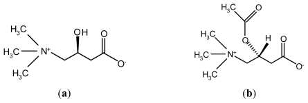
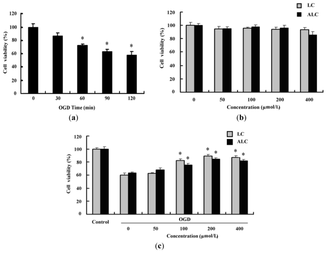
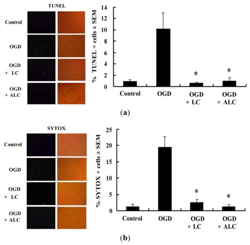
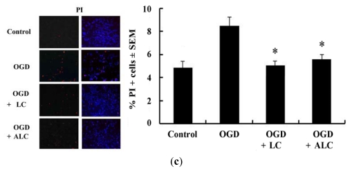
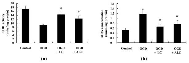
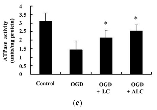
The therapeutic effect of stroke is hampered by the lack of neuroprotective drugs against ischemic insults beyond the acute phase. Carnitine plays important roles in mitochondrial metabolism and in modulating the ratio of coenzyme A (CoA)/acyl-CoA. Here, we investigate the neuroprotective effects of l-carnitine (LC) and Acetyl-l-carnitine (ALC) pre-treatment on ischemic insults under the same experimental conditions. We used a transient middle cerebral artery occlusion (MCAO) model to evaluate the protective roles of LC and ALC in acute focal cerebral ischemia in vivo and to understand the possible mechanisms using model of PC12 cell cultures in vitro. Results showed that ALC, but not LC, decreased infarction size in SD rats after MCAO in vivo. However, both LC and ALC pretreatment reduced oxygen-glucose deprivation (OGD)-induced cell injury and decreased OGD-induced cell apoptosis and death in vitro; at the same time, both of them increased the activities of super oxide dismutase (SOD) and ATPase, and decreased the concentration of malondialdehyde (MDA) in vitro. Thus, our findings suggested that LC and ALC pre-treatment are highly effective in the prevention of neuronal cell against ischemic injury in vitro, however, only ALC has the protective effect on neuronal cell injury after ischemia in vivo.
Keywords: Acetyl-l-carnitine; ischemia; l-carnitine; neuroprotection; oxygen-glucose deprivation.
Figures

Similar articles
-
Neuroprotective Effects of Acetyl-L-Carnitine Against Oxygen-Glucose Deprivation-Induced Neural Stem Cell Death.Mol Neurobiol. 2016 Dec;53(10):6644-6652. doi: 10.1007/s12035-015-9563-x. Epub 2015 Dec 8. Mol Neurobiol. 2016. PMID: 26643543
-
Acetyl-l-carnitine restores synaptic transmission and enhances the inducibility of stable LTP after oxygen-glucose deprivation.Neuroscience. 2016 Sep 22;332:203-11. doi: 10.1016/j.neuroscience.2016.06.046. Epub 2016 Jul 1. Neuroscience. 2016. PMID: 27378558
-
Neuroprotective effect of ginkgolide K against acute ischemic stroke on middle cerebral ischemia occlusion in rats.J Nat Med. 2012 Jan;66(1):25-31. doi: 10.1007/s11418-011-0545-7. Epub 2011 May 25. J Nat Med. 2012. PMID: 21611909
-
Mechanisms of ischemic neuroprotection by acetyl-L-carnitine.Ann N Y Acad Sci. 2005 Aug;1053:153-61. doi: 10.1196/annals.1344.013. Ann N Y Acad Sci. 2005. PMID: 16179519 Free PMC article. Review.
-
The neurobiology of acetyl-L-carnitine.Front Biosci (Landmark Ed). 2016 Jun 1;21(7):1314-29. doi: 10.2741/4459. Front Biosci (Landmark Ed). 2016. PMID: 27100509 Review.
Cited by
-
CSPGs inhibit axon branching by impairing mitochondria-dependent regulation of actin dynamics and axonal translation.Dev Neurobiol. 2017 Apr;77(4):454-473. doi: 10.1002/dneu.22420. Epub 2016 Aug 2. Dev Neurobiol. 2017. PMID: 27429169 Free PMC article.
-
Organic and Peptidyl Constituents of Snake Venoms: The Picture Is Vastly More Complex Than We Imagined.Toxins (Basel). 2018 Sep 26;10(10):392. doi: 10.3390/toxins10100392. Toxins (Basel). 2018. PMID: 30261630 Free PMC article.
-
Emerging antioxidant therapies in Friedreich's ataxia.Front Pharmacol. 2024 Feb 6;15:1359618. doi: 10.3389/fphar.2024.1359618. eCollection 2024. Front Pharmacol. 2024. PMID: 38379897 Free PMC article. Review.
-
Assessment of L-carnitine effectiveness on carpal tunnel syndrome.Curr J Neurol. 2022 Jul 6;21(3):162-169. doi: 10.18502/cjn.v21i3.11109. Curr J Neurol. 2022. PMID: 38011355 Free PMC article.
-
Neuroprotective Effects of Acetyl-L-Carnitine Against Oxygen-Glucose Deprivation-Induced Neural Stem Cell Death.Mol Neurobiol. 2016 Dec;53(10):6644-6652. doi: 10.1007/s12035-015-9563-x. Epub 2015 Dec 8. Mol Neurobiol. 2016. PMID: 26643543
References
-
- Murray C.J., Lopez A.D. Mortality by cause for eight regions of the world: Global burden of disease study. Lancet. 1997;349:1269–1276. - PubMed
-
- Deshpande J.K., Siesjo B.K., Wieloch T. Calcium accumulation and neuronal damage in the rat hippocampus following cerebral ischemia. J. Cereb. Blood Flow Metab. 1987;7:89–95. - PubMed
-
- MacManus J.P., Buchan A.M., Hill I.E., Rasquinha I., Preston E. Global ischemia can cause DNA fragmentation indicative of apoptosis in rat brain. Neurosci. Lett. 1993;164:89–92. - PubMed
-
- Leist M., Jaattela M. Four deaths and a funeral: From caspases to alternative mechanisms. Nat. Rev. Mol. Cell Biol. 2001;2:589–598. - PubMed
-
- Picconi B., Barone I., Pisani A., Nicolai R., Benatti P., Bernardi G., Calvani M., Calabresi P. Acetyl-l-carnitine protects striatal neurons against in vitro ischemia: The role of endogenous acetylcholine. Neuropharmacology. 2006;50:917–923. - PubMed
Publication types
MeSH terms
Substances
LinkOut - more resources
Full Text Sources
Other Literature Sources
