

doi: 10.3390/s90604407.
Epub 2009 Jun 5.
Antibody-based sensors: principles, problems and potential for detection of pathogens and associated toxins
Affiliations
- PMID: 22408533
- PMCID: PMC3291918
- DOI: 10.3390/s90604407
Item in Clipboard
Antibody-based sensors: principles, problems and potential for detection of pathogens and associated toxins
Sensors (Basel).
2009.
Abstract
Antibody-based sensors permit the rapid and sensitive analysis of a range of pathogens and associated toxins. A critical assessment of the implementation of such formats is provided, with reference to their principles, problems and potential for 'on-site' analysis. Particular emphasis is placed on the detection of foodborne bacterial pathogens, such as Escherichia coli and Listeria monocytogenes, and additional examples relating to the monitoring of fungal pathogens, viruses, mycotoxins, marine toxins and parasites are also provided.
Keywords: antibody; assay development; biosensor; electrochemical; pathogen; surface-plasmon resonance.
Figures
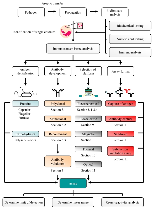
Strategy for pathogen detection.
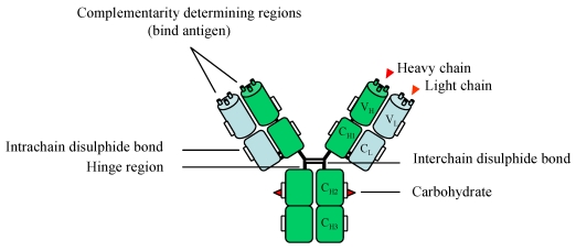
A schematic representation of an IgG antibody comprising of two heavy (green) and light (blue) chains. Carbohydrate elements are attached via the asparagine 297 amino acid residue. A more in-depth discussion of antibody glycosylation is provided in reference [30]. Key: VH – variable heavy, VL – variable light, CH – constant heavy, CL – constant light.
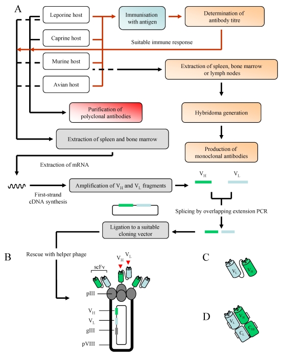
An overview of monoclonal, polyclonal and recombinant antibody production [A]. Immunisation-related stages are represented by a red line, with those involving antibody production shown in black. A more in-depth discussion of the generation of recombinant antibodies, inclusive of Fab fragments, can be found in reference [38]. Additional hosts may also be used for antibody production, including camels (camelid), sheep (ovine) and pigs (porcine). A filamentous phage displaying scFv antibody fragments [B] and two recombinant antibody fragments, the scFv [C] and Fab [D], are also illustrated. Key: pIII/pVIII – protein 3/8, VH – variable heavy, VL – variable light.
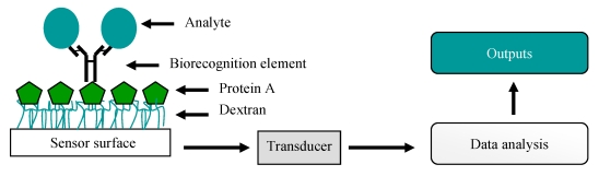
A simple representation of a biosensor. Here, a full-length antibody is captured on protein A immobilised on a carboxymethylated dextran-coated sensor surface and is used for the capture of an analyte. This interaction produces a specific physicochemical change, such as a change in mass, temperature or electrical potential. This is then converted (via a transducer) to a signal which the user can interpret.
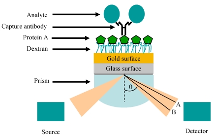
Representation of the SPR phenomenon, showing the Kretschmann prism arrangement originally proposed in references [86] and [87]. For illustrative purposes, a protein-A (green hexagon)-captured IgG antibody is shown on a carboxymethylated dextran (CM5) sensor surface. The mass change introduced by the binding of an analyte of interest (blue circle) is shown as a change in refractive index (A to B) which can be determined through the use of dedicated software.
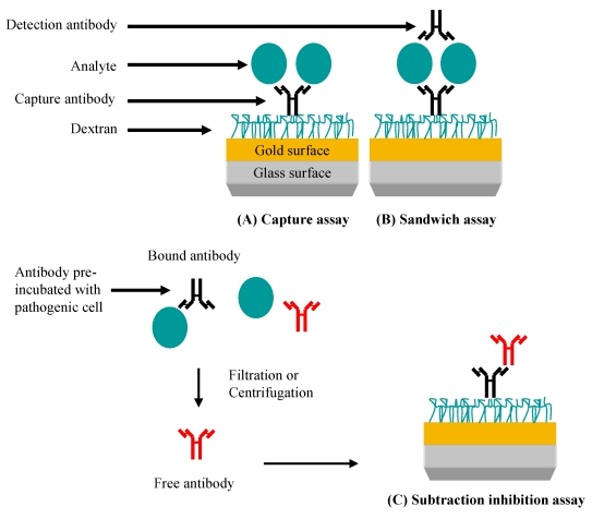
SPR-based assays for pathogen detection. (A) Specific antibody is immobilised and is used to capture the pathogen leading to a signal. (B) Pathogen or pathogen-related antigen is captured. Specificity is conferred by the binding of a second antibody. (C) Specific antibody reacts with the pathogen or pathogen-related antigen. Non-bound (free) antibody is isolated and detected when bound to an immobilised antibody (normally an anti-species antibody) on the chip. In this case, the signal generated is inversely proportional to the pathogen concentration.
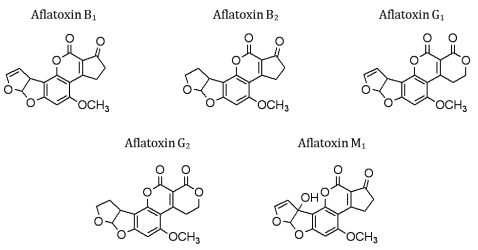
Structures of commonly encountered aflatoxins.
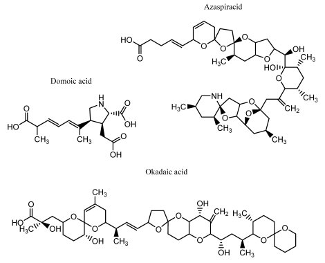
Structures of commonly encountered phycotoxins.
References
-
- Gracias K.S., McKillip J.L. A review of conventional detection and enumeration methods for pathogenic bacteria in food. Can. J. Microbiol. 2004;50:883–890. - PubMed
-
- Bhunia A.K. Biosensors and bio-based methods for the separation and detection of foodborne pathogens. Adv. Food Nutr. Res. 2008;54:1–44. - PubMed
-
- Leonard P., Hearty S., Brennan J., Dunne L., Quinn J., Chakraborty T., O'Kennedy R. Advances in biosensors for detection of pathogens in food and water. Enzyme Microb. Tech. 2003;32:3–13.
-
- Available online: www.oxoid.com/UK/blue/orgbrowse/orgbrowse.asp, Accession date: May 6, 2009
LinkOut - more resources
Full Text Sources
Other Literature Sources
