. 2012 Mar 6:6:7.
doi: 10.3389/fnana.2012.00007. eCollection 2012 Jan 7.

Developmental disorders of the midbrain and hindbrain

Affiliations

Developmental disorders of the midbrain and hindbrain

A James Barkovich. Front Neuroanat. .

Abstract

Malformations of the midbrain (MB) and hindbrain (HB) have become topics of considerable interest in the neurology and neuroscience literature in recent years. The combined advances of imaging and molecular biology have improved analyses of structures in these areas of the central nervous system, while advances in genetics have made it clear that malformations of these structures are often associated with dysfunction or malformation of other organ systems. This review focuses upon the importance of communication between clinical researchers and basic scientists in the advancement of knowledge of this group of disorders. Disorders of anteroposterior (AP) patterning, cerebellar hypoplasias, disorders associated with defects of the pial limiting membrane (cobblestone cortex), disorders of the Reelin pathway, and disorders of the primary cilium/basal body organelle (molar tooth malformations) are the main focus of the review.

Keywords: cerebellum; hindbrain; malformations; midbrain.


Figures
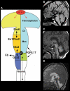
Figure 1
Figure 1
Disorders of AP patterning in the brain stem.(A) Anteroposterior patterning and the Isthmus Organizer. Regionalization of the brain starts with the formation of patterning centers that secrete signaling molecules such as the fibroblast growth factors (FGFs). Fgf8 and Fgr17 are important signaling molecules at both the anterior forebrain and the MB-HB junction. In the forebrain, it helps to direct formation of the prefrontal cortex and other rostral structures by inducing cells to secrete the transcription factor Pax6. At the MB-HB junction, the patterning center known as the isthmus organizer (IsO) is localized and induced to secrete Fgf8 and Fgf17 by the interaction of transcription factor Gbx2 from the rhombencephalon and Otx2 from the caudal mesencephalon. The secretion of Fgf8 and Fgf17 then induces further changes crucial to formation of the MB-HB junction and the formation of the cerebellum. The junction of the diencephalon (di) and mesencephalon (mes) is directed by the interaction of Pax6 from the diencephalon and En1/Pax2 from the rostral mesencephalon. Repression of Otx2 expression by FGF8 induces Gbx2 formation to establish the location of the MB-HB junction and can affect cerebellar formation, as the cerebellum forms from the most rostral portion of the HB. Similarly, alterations of Pax6 or En1/Pax2 will alter the location of the diencephalic-mesencephalic junction. (Adapted from Barkovich and Raybaud, 2012). (B) Sagittal T1 weighted image shows a short MB, long pons, and large superior vermis (black arrows), suggesting an abnormality of anteroposterior patterning with rostral misplacement of the Isthmus Organizer. In addition, the patient has agenesis of the corpus callosum. (C) Sagittal T1 weighted image shows a slightly small pons and a short, thick medulla (white arrows) with an abnormal pontomedullary transition. This is postulated to result from mixed gains and losses of rhombomere expression in the developing rhombencephalon or potentially a segmental shift of rhombomeres. (D) Sagittal T2 weighted image shows a very elongated MB (white arrows) with small, short pons (black arrow), and small cerebellar vermis, suggesting caudal displacement of the Isthmus Organizer due to abnormal anterioposterior patterning from overexpression of Otx2.
Figure 2
Figure 2
The concept and range of cerebellum/posterior fossa disorders in the “Dandy–Walker spectrum.”(A) Illustration of developing fourth ventricle/cerebellum and the impact of impaired egress of CSF. During normal development, the wall fourth ventricle normally thins, and the foramen of Magendie forms, in the midline. If the leptomeninges are abnormal, the cerebellum may be small and the outflow foramina of the fourth ventricle may not form. The fourth ventricle expands posteriorly (small black arrows) and superiorly, pushing the small cerebellar vermis (Cb) counterclockwise (large black arrow); the posterior fossa then enlarges. This combination of findings creates the classic Dandy–Walker malformation. Small black PF signifies pontine flexure, small black MF signifies mesencephalic flesure, large black P signifies prosencephalon, large black M signifies mesencephalon, large black R signifies rhombencephalon. (B) Classic Dandy–Walker malformation. Sagittal T2 weighted image shows the classic appearance with a small vermis (black arrow), rotated counterclockwise with abnormal foliation. The surrounding CSF spaces are markedly enlarged with abnormally enlarged posterior fossa and elevation of the tentorium cerebelli and the torcular Herophili. (C) Mega cisterna magna is a condition in which a collection of CSF in an enlarged cisterna magna (C) expands the posterior fossa but the midbrain and hindbrain are normal. It is seen in patients with gene mutations that, in siblings, cause the classic Dandy–Walker malformation. (D) Blake pouch cyst is a condition in which the ependymal wall of the fourth ventricle stretches out through the foramen of Magendie and causes enlargement of the foramen with mild rotation of the vermis (black arrow). It is considered by some to be an incidental finding and by others to be a mild form of the Dandy–Walker malformation.
Figure 3
Figure 3
Array of findings in the midbrain and hindbrain of cobblestone malformations (formerly called Lissencephaly type II). Diagram (A) shows neuron (n) guided by normal radial glial cell (NL RGC) on the left, coursing from ependyma to an intact pial limiting membrane (Pial LM), where it attaches via a bridge made by beta dystroglycan, alpha dystroglycan, or GPR56, which attach to laminin-2 or collagen IV in the Pial LM. In the center and on the right, gaps are seen in the Pial LM; the RGCs do not attach properly due to defects of alpha dystroglycan or GPR56 in the leading process of the RGC, to laminin, or collagen IV, respectively, in the Pial LM. Neurons either detach prematurely or overmigrate through the gaps into the subarachnoid space. A relatively mild cerebellar anomaly is shown in the muscle-eye-brain phenotype shown in (B) and (C). Although the vermis is small and dysmorphic, the hemispheres have nearly normal foliation. A few small cysts are present (white arrows in B and C). The pons contains a midline cleft (black arrow in C). The midbrain tectum is large and smooth due to transpial migration of cells. A coronal image through the cerebrum (D) shows moderate ventricular enlargement, abnormal hyperintensity of subcortical, and deep white matter, and abnormal sulcation over the convexities; note that the cortex in this region (white arrows) is abnormally thick and seems to be formed of radially oriented bands of neurons. A much more severe Walker–Warburg phenotype is shown in (E) and (F). The sagittal image (E) shows a thin brain stem with a large kink in the mid pons (black arrowhead), resembling a persistent pontine flexure. The MB tectum is very large and rounded (large black arrow). Only a small vermis (small black arrow) is present. Massive hydrocephalus can be seen, as can a small occipital cephalocele (white arrow). The axial image (F) shows an extremely small, dysmorphic cerebellum with no vermis and many cysts within the irregular cortex. Both ocular globes are anomalous.
Figure 4
Figure 4
Array of findings in cerebrum and cerebellum of disorders of the Reelin pathway. The most severe situation, with severe RELN depletion results in a very small vermis (white arrow in A), small, smooth cerebellar hemispheres (black arrows in B), and a thick, pachygyric cerebral cortex (B). VLDLR mutation results in a less severe cerebral dysgenesis (cortex is thinner and more sulci are present) but the cerebellum is quite severely affected, both hypoplastic and smooth (C). Severe cerebral but less severe cerebellar involvement (note that the cerebellum is larger and cortex is less smooth) (D) can be seen, but the precise mechanism/mutation that results in this appearance is not known.
Figure 5
Figure 5
Neuroimaging findings in molar tooth malformations. The characteristic imaging findings of a small vermis (small white arrows, A,B) and narrow isthmus (small white arrowhead, A,B) are identified on sagittal images. A tuber cinereum hamartoma is seen in (B). The variable appearances of the “molar tooth,” resulting from the large, horizontal superior cerebellar peduncles, are shown (white arrows in C, D, and E).

References

    1. Ackroyd M. R., Whitmore C., Prior S., Kaluarachchi M., Nikolic M., Mayer U., Muntoni F., Brown S. C. (2011). Fukutin-related protein alters the deposition of laminin in the eye and brain. J. Neurosci. 31, 12927–12935 10.1523/JNEUROSCI.2301-11.2011 - DOI - PMC - PubMed
    1. Acosta F. L. Jr., Binder D. K., Barkovich A. J., Frieden I. J., Gupta N. (2005). Neurocutaneous melanosis presenting with hydrocephalus. Case report and review of the literature. J. Neurosurg. 102(Suppl. 1), 96–100 10.3171/ped.2005.102.1.0096 - DOI - PubMed
    1. Aida N., Yagishita A., Takada K., Katsumata Y. (1994). Cerebellar MR in Fukuyama congenital muscular dystrophy: polymicrogyria with cystic lesions. AJNR. Am. J. Neuroradiol. 15, 1755–1759 - PMC - PubMed
    1. Aldinger K. A., Lehmann O. J., Hudgins L., Chizhikov V. V., Bassuk A. G., Ades L. C., Krantz I. D., Dobyns W. B., Millen K. J. (2009). FOXC1 is required for normal cerebellar development and is a major contributor to chromosome 6p25.3 Dandy-Walker malformation. Nat. Genet. 41, 1037–1042 10.1038/ng.422 - DOI - PMC - PubMed
    1. Aranda-Orgillés B., Trockenbacher A., Winter J., Aigner J., Köhler A., Jastrzebska E., Stahl J., Müller E.-C., Otto A., Wanker E., Schneider R., Schweiger S. (2008). The Opitz syndrome gene product MID1 assembles a microtubule-associated ribonucleoprotein complex. Hum. Genet. 123, 163–176 10.1007/s00439-007-0456-6 - DOI - PMC - PubMed