Review
. 2012 Jun;80(6):1948-57.
doi: 10.1128/IAI.06014-11. Epub 2012 Mar 12.

Membrane vesicle release in bacteria, eukaryotes, and archaea: a conserved yet underappreciated aspect of microbial life

Affiliations
Review

Membrane vesicle release in bacteria, eukaryotes, and archaea: a conserved yet underappreciated aspect of microbial life

Brooke L Deatherage et al. Infect Immun. 2012 Jun.

Abstract

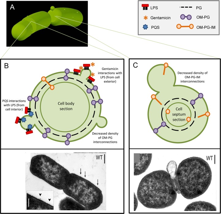
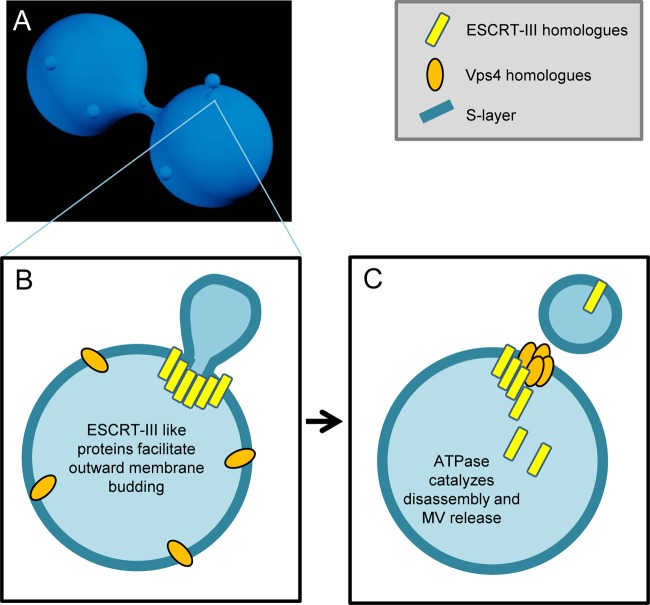
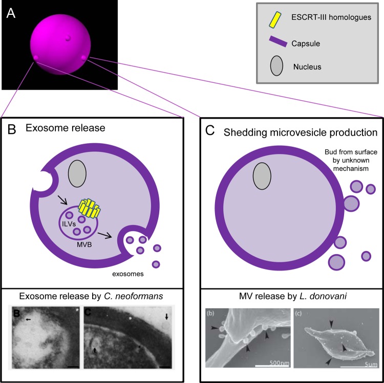
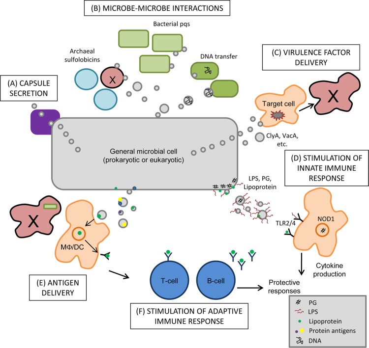
Interaction of microbes with their environment depends on features of the dynamic microbial surface throughout cell growth and division. Surface modifications, whether used to acquire nutrients, defend against other microbes, or resist the pressures of a host immune system, facilitate adaptation to unique surroundings. The release of bioactive membrane vesicles (MVs) from the cell surface is conserved across microbial life, in bacteria, archaea, fungi, and parasites. MV production occurs not only in vitro but also in vivo during infection, underscoring the influence of these surface organelles in microbial physiology and pathogenesis through delivery of enzymes, toxins, communication signals, and antigens recognized by the innate and adaptive immune systems. Derived from a variety of organisms that span kingdoms of life and called by several names (membrane vesicles, outer membrane vesicles [OMVs], exosomes, shedding microvesicles, etc.), the conserved functions and mechanistic strategies of MV release are similar, including the use of ESCRT proteins and ESCRT protein homologues to facilitate these processes in archaea and eukaryotic microbes. Although forms of MV release by different organisms share similar visual, mechanistic, and functional features, there has been little comparison across microbial life. This underappreciated conservation of vesicle release, and the resulting functional impact throughout the tree of life, explored in this review, stresses the importance of vesicle-mediated processes throughout biology.


Figures

Fig 1
Fig 1
MV production by Bacteria. (A) Gram-negative bacteria release surface-derived membrane vesicles (MVs) at division septa and along the length of the cell body. (B) Upper panel: proteins harboring domains that link the outer membrane (OM) to the peptidoglycan layer (PG) minimize MV release along the cell body; temporary disruption of these OM-PG interactions results in MV release. Membrane-active antibiotics (such as gentamicin) and signaling molecules (such as pqs) interact with the membrane surface to induce MV release. Lower panel: transmission electron micrograph (TEM) of wild-type S. Typhimurium with inset showing small MV release along the cell body. (Reprinted from reference with permission of the publisher, John Wiley and Sons.) (C) Upper panel: at the constricted division septum, temporary dissociation of OM-PG-IM protein complexes spanning the OM and inner membrane (IM) occurs, facilitating the release of a large MV before completing cell division. Lower panel: TEM of wild-type S. Typhimurium releasing a septal MV. (Reprinted from reference with permission of the publisher, John Wiley and Sons.)
Fig 2
Fig 2
MV production by Archaea. (A) Archaea release MVs that are derived from the cell surface, similar to the process in Bacteria. (B) This process is facilitated through the coordinated action of ESCRT-III homologue proteins (yellow), conserved in archaeal and eukaryotic life. ESCRT-III homologues, known for membrane scission capabilities, are directed to surround the site of nascent MV formation and induce the outward protrusion of the membrane, including the archaeal S-layer. (C) Vps4 homologue ATPases then catalyze the disassembly of ESCRT-III homologues, and MV release occurs.
Fig 3
Fig 3
MV production by eukaryotic microbes. (A) Eukaryotic microbes, including fungi (shown here) and parasites, release MVs at the cell surface, although these MVs may be derived from multiple sources. (B) Upper panel: exosome release is a conserved process in eukaryotic microbes. An endosome is created in the cytosol, which traffics through the microbial cell. In transit, the ESCRT-III homologues (also conserved in Archaea and higher eukaryotes) induce formation of intraluminal vesicles (ILVs), creating multivesicular bodies (MVBs). MVBs fuse to the cell surface and release the vesicular content as exosomes. Lower panel: electron micrograph demonstrating release of exosomes by the fungus Cryptococcus neoformans. (Reprinted from reference with permission.) (C) An additional pathway for MV release in eukaryotic microbes exists, appearing to produce surface-derived MVs reminiscent of those in Bacteria and Archaea. In this process, for which mechanistic details are as yet unknown, shedding microvesicles bud directly from the cell surface. Lower panel: electron micrograph demonstrating MV release by the parasite Leishmania donovani. (Reprinted from reference with permission of the publisher, BioMed Central.)
Fig 4
Fig 4
Biological impact of MV release. MVs, originating from bacteria, fungi, archaea, or parasites, possess many functions in microbial physiology and pathogenesis. MVs promote the secretion of capsular polysaccharide to the cell surface (A) and are utilized for cell-cell communication between Bacteria (release of quorum-sensing molecules and transfer of DNA) and Archaea (secretion of antimicrobials) (B). (C) For pathogenic microbes, the role of MVs in vivo is likely multifaceted, including the ability to directly deliver virulence factors, such as toxins, to target host cells. (D) Natural microbial structures present in MVs can act to stimulate the innate immune system through activation of TLRs (via LPS and/or lipoprotein sensing) and NLRs (via peptidoglycan detection). (E) These antigenic structures contained in MVs likely interact with antigen-presenting cells (APC) when released in vivo during infection, facilitating the presentation of MV antigens in cases where APC may be impaired by cytotoxic organisms. (F) Antigen presentation can lead to the stimulation of adaptive immune responses, triggering T-cell and B-cell responses that are directed toward MV antigens. The ways in which MVs are utilized by microbes during growth (and during infection in the case of pathogenic organisms) are complex and they underscore the importance of these structures for all microbial life.

References

    1. Abadi J, Pirofski L. 1999. Antibodies reactive with the cryptococcal capsular polysaccharide glucuronoxylomannan are present in sera from children with and without human immunodeficiency virus infection. J. Infect. Dis. 180:915–919 - PubMed
    1. Alaniz RC, Deatherage BL, Lara JC, Cookson BT. 2007. Membrane vesicles are immunogenic facsimiles of Salmonella typhimurium that potently activate dendritic cells, prime B and T cell responses, and stimulate protective immunity in vivo. J. Immunol. 179:7692–7701 - PubMed
    1. Albuquerque PC, et al. 2008. Vesicular transport in Histoplasma capsulatum: an effective mechanism for trans-cell wall transfer of proteins and lipids in ascomycetes. Cell. Microbiol. 10:1695–1710 - PMC - PubMed
    1. Bergman MA, et al. 2005. CD4+ T cells and toll-like receptors recognize Salmonella antigens expressed in bacterial surface organelles. Infect. Immun. 73:1350–1356 - PMC - PubMed
    1. Bernadac A, Gavioli M, Lazzaroni JC, Raina S, Lloubes R. 1998. Escherichia coli tol-pal mutants form outer membrane vesicles. J. Bacteriol. 180:4872–4878 - PMC - PubMed

Publication types

LinkOut - more resources