Review
. 2012 Jun;37(6):220-9.
doi: 10.1016/j.tibs.2012.01.003. Epub 2012 Mar 10.

Molecular underpinnings of centromere identity and maintenance

Affiliations
Review

Molecular underpinnings of centromere identity and maintenance

Nikolina Sekulic et al. Trends Biochem Sci. 2012 Jun.

Abstract

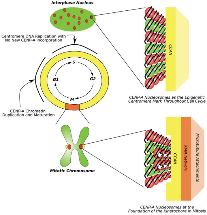
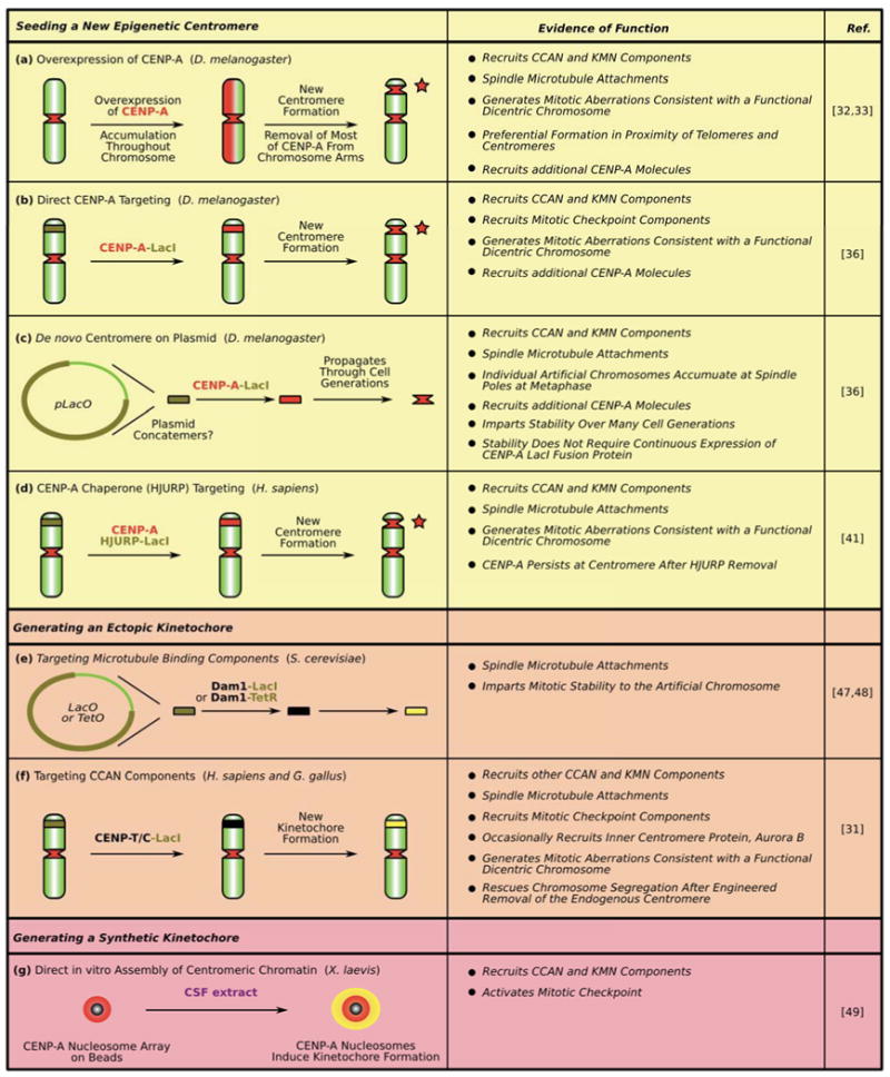
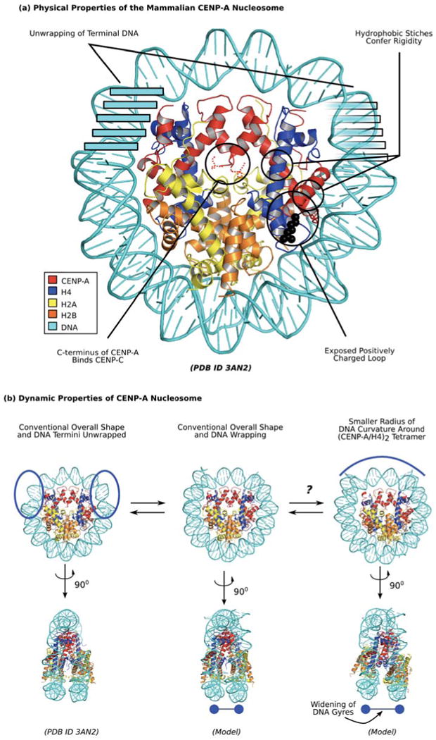
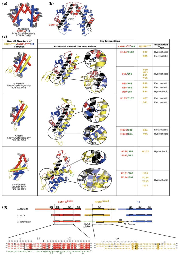
Centromeres direct faithful chromosome inheritance at cell division but are not defined by a conserved DNA sequence. Instead, a specialized form of chromatin containing the histone H3 variant, CENP-A, epigenetically specifies centromere location. We discuss current models where CENP-A serves as the marker for the centromere during the entire cell cycle in addition to generating the foundational chromatin for the kinetochore in mitosis. Recent elegant experiments have indicated that engineered arrays of CENP-A-containing nucleosomes are sufficient to serve as the site of kinetochore formation and for seeding centromeric chromatin that self-propagates through cell generations. Finally, recent structural and dynamic studies of CENP-A-containing histone complexes - before and after assembly into nucleosomes - provide models to explain underlying molecular mechanisms at the centromere.


Figures

Figure 1
Figure 1. Epigenetic marking and kinetochore nucleating functions of CENP-A-containing nucleosomes
Diagram showing that CENP-A (red dots in the green interphase nucleus and on the mitotic chromosome) marks centromere location throughout the cell cycle. Two key points during interphase that are currently under investigation are S-phase when centromere DNA is replicated but no new CENP-A is deposited and early G1 when new CENP-A protein incorporates at centromeres and results in doubling the number of CENP-A nucleosomes. Throughout the cell cycle, CENP-A nucleosomes are associated with the constitutive centromere-associated protein network (CCAN) (represented by a yellow rectangle). In mitosis, CENP-A nucleosomes are (re)organized on the face of the chromosome. Connections are made between the CCAN and the so-called KMN network (orange rectangle) that is directly involved in attaching to spindle microtubules [82]. As detailed in the text, one untested possibility is that specialized and somehow modified subset of CENP-A nucleosomes (denoted with white stars) serve as the key connection point, linking the chromosome to a spindle microtubule through the CCAN and KMN network.
Figure 2
Figure 2. Summary of various experimental approaches used to seed a new epignetic centromere (a-d), to generate an ectopic kinetochore (e-f) and a synthetic kinetochore (g)
See text for details. Dark green segments with black line boundaries on chromosome arms denote the location of integrated LacO arrays. Red stars denote location of the new centromere in panels a, b and d. Yellow denotes kinetochore activity in panels e-g.
Figure 3
Figure 3. Unique physical properties of the mammalian CENP-A-containing nucleosome
(a) Distinguished physical properties of CENP-A nucleosome are highlighted in black circles. Clockwise from the top left, these include transient unwrapping/flexibility of the final helical turn of DNA at each nucleosome terminus [14,57,58], hydrophobic stitches that rigidify the CENP-A/H4 interface [13,50,54,56], a bulged loop L1 that is of opposite charge as on H3 in the conventional nucleosome [13,14], and the unstructured C-terminus that mediates recognition of CENP-A nucleosomes by CENP-C [83]. (b) Dynamic properties of CENP-A nucleosome indicate potential sampling between distinct conformations. Mammalian CENP-A containing nucleosomes wrap ∼145 bp of DNA at steady-state (center; [13,58]), but sample unwrapping at the terminal DNA to a greater extent than conventional nucleosomes with canonical H3 [57,58]. The crystal form of the nucleosome includes only 120 bp of DNA that is visible in the structure (left; [14]), supporting the notion of increased flexibility at nucleosome termini. Another potential alternate form under investigation is one where rotation at the CENP-A/CENP-A is maintained as in the isolated sub-nucleosomal (CENP-A/H4)2 heterotetramer [13], leading to a smaller radius of curvature of DNA wrapping near the nucleosomal dyad (right, top) and wider spacing of H2A/H2B heterodimers and the two strands of nucleosomal DNA (right, bottom).
Figure 4
Figure 4. Structures of HJURPScm3/CENP-ACse4/H4 ternary complexes
(a) The sub-nucleosomal (CENP-A/H4)2 structure [13] is shown with cylindrical helices as a reference point for how CENP-A and H4 are affected by binding to HJURPScm3. Positively charged residues making DNA contacts [14] are depicted as grey spheres. (b) Ribbon diagram of CENP-A/H4 tetramer (PDB ID 3NQJ) with the CATD of CENP-A colored black [13] shown for easier comparison with complexes in panel c. (c) Structure of HJURPScm3 (yellow)/CENP-ACse4 (red)/H4 (blue) complexes [62,72,73] with cylindrical helices (right column) and details of specific interactions in each of the structures (middle and right columns). The middle column includes ribbon diagrams of complexes with sites of specific interactions circled in black and accompanying enlargements. For easier comparison between each other and the tetramer in panel b, all structures were overlaid using residues within α2 helix of CENP-A (labeled black). (d) Linear diagram of constructs used in the three HJURPScm3/CENP-ACse4/H4 ternary complex structural studies. In constructs used for crystal structures, the residues present in the construct but lacking order in the corresponding structure are made transparent. The shared contact surfaces in each structure are contained within the dashed boxed regions of CENP-ACse4 (red) and HJURPScm3 (yellow), with α-helices indicated by small solid boxes and β-strands indicated by arrows. The S. cerevisiae study used a single-chain fused polypeptide with a 6 a.a. linker sequence between CENP-ACse4 and HJURPScm3 (green) and no linker between HJURPScm3 and H4. Sequence overlays are presented underneath the scheme with conserved residues between all structures labeled with boxes of solid colors (red for CENP-ACse4 and yellow for HJURPScm3). The CATD of CENP-A is boxed with a black rectangle. The interacting residues (listed in panel c) proposed as determinants of specificity between CENP-ACse4 and HJURPScm3 are shown in black. For easier analysis, the corresponding region of human histone H3 sequence (green) is placed underneath CENP-ACse4 sequences. Six residues differ between human H3.1 and fungal H3 proteins and the matching amino acids in fungal H3 are shown below the human H3 sequence.

References

    1. Cleveland DW, et al. Centromeres and kinetochores: from epigenetics to mitotic checkpoint signaling. Cell. 2003;112:407–421. - PubMed
    1. Allshire RC, Karpen GH. Epigenetic regulation of centromeric chromatin: old dogs, new tricks? Nat Rev Genet. 2008;9:923–937. - PMC - PubMed
    1. Eichler EE. Repetitive conundrums of centromere structure and function. Hum Mol Genet. 1999;8:151–155. - PubMed
    1. Henikoff S, et al. The centromere paradox: stable inheritance with rapidly evolving DNA. Science. 2001;293:1098–1102. - PubMed
    1. Murphy WJ, et al. Dynamics of mammalian chromosome evolution inferred from multispecies comparative maps. Science. 2005;309:613–617. - PubMed

Publication types

MeSH terms

LinkOut - more resources