. 2012 May;241(5):852-62.
doi: 10.1002/dvdy.23771. Epub 2012 Mar 23.

Inhibitory morphogens and monopodial branching of the embryonic chicken lung

Affiliations

Inhibitory morphogens and monopodial branching of the embryonic chicken lung

Jason P Gleghorn et al. Dev Dyn. 2012 May.

Abstract

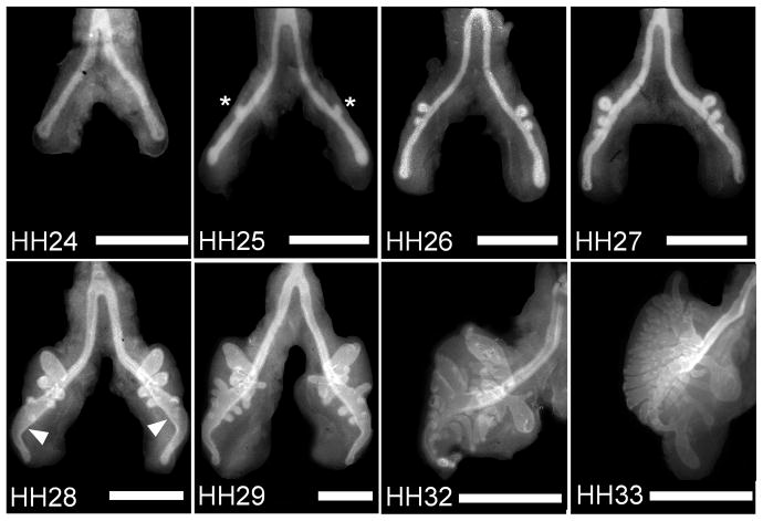
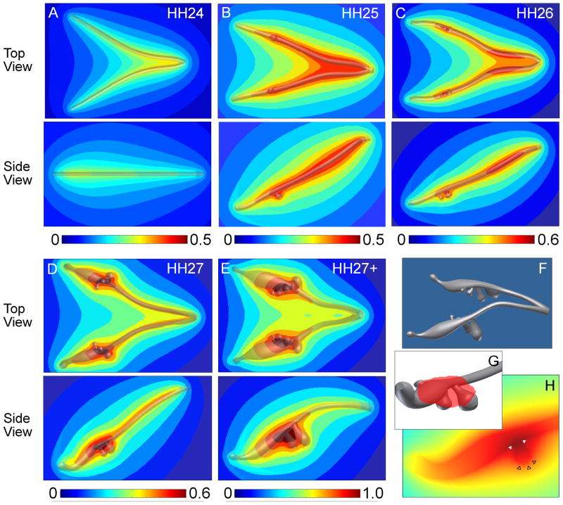
Background: Branching morphogenesis generates a diverse array of epithelial patterns, including dichotomous and monopodial geometries. Dichotomous branching can be instructed by concentration gradients of epithelial-derived inhibitory morphogens, including transforming growth factor-β (TGFβ), which is responsible for ramification of the pubertal mammary gland. Here, we investigated the role of autocrine inhibitory morphogens in monopodial branching morphogenesis of the embryonic chicken lung.

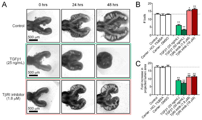
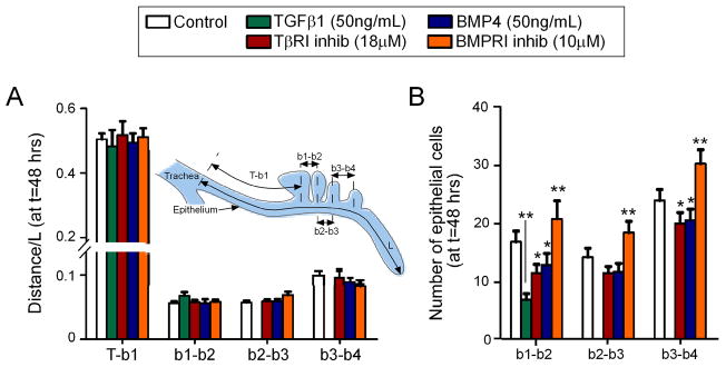
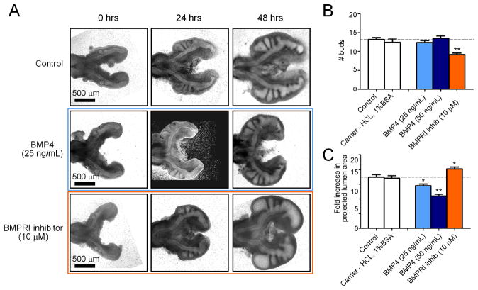
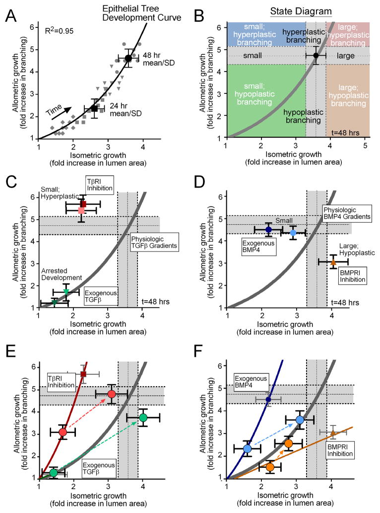
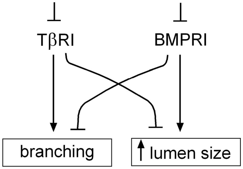
Results: Computational modeling and experiments using cultured organ explants each separately revealed that monopodial branching patterns cannot be specified by a single epithelial-derived autocrine morphogen gradient. Instead, signaling by means of TGFβ1 and bone morphogenetic protein-4 (BMP4) differentially affect the rates of branching and growth of the airways. Allometric analysis revealed that development of the epithelial tree obeys power-law dynamics; TGFβ1 and BMP4 have distinct but reversible effects on the scaling coefficient of the power law.

Conclusions: These data suggest that although autocrine inhibition cannot specify monopodial branching, inhibitory morphogens define the dynamics of lung morphogenesis.


Figures

Figure 1
Figure 1
Development of embryonic chicken airways over time. Immunofluorescence images of whole mount staining for E-cadherin in chicken lungs (developmental stages HH24-33) reveal a monopodial branching pattern. Asterisks denote examples of emerging secondary bronchi; arrowheads indicate the vestibulum. Scale bars, 500 μm.
Figure 2
Figure 2
Computational analysis of morphogen concentration and distribution in developing chicken lungs. (A–E) Predicted concentration profiles of a hypothetical morphogen uniformly secreted from the epithelium at various developmental stages. The concentration profiles are normalized to the highest value calculated for the HH27+ model, with red indicating areas of highest relative concentration and blue representing regions of low morphogen concentration. (F) An example solid model (HH27+) used for FEM analysis that was created from dimensions of E-cadherin-stained lung explants. (G) 3D solid model representation of the region of highest morphogen concentration represented by the red shading and (H) cross-section plot through a bud plotting morphogen concentration, which illustrates that the bud stalk and branch point have a local maximum (solid white triangles) whereas the bud tip has a local minimum (empty black triangles) in predicted morphogen concentration.
Figure 3
Figure 3
Effect of disrupting the endogenous TGFβ concentration ex vivo. (A) Representative brightfield images of lungs cultured over 48 hours with recombinant TGFβ1, TβRI inhibitor, or control. Images are for the same explants tracked over time. Quantitative measures of bud enumeration (B) and fold increase in projected lumen area (C) of treated lungs after 48 hours of culture. Data +/− SEM (n>10), * p<0.05, ** p<0.001 relative to controls.
Figure 4
Figure 4
Relative position of secondary bronchi along the primary bronchus. (A) Distance of each bud (b1, b2, b3, b4) normalized to the length of the primary bronchus (L), as measured from the tracheal bifurcation (T). (B) Average number of epithelial cells along the primary bronchus between subsequent secondary bronchi. Data +/− SEM (n>10), * p<0.05, ** p<0.001 relative to controls.
Figure 5
Figure 5
Effect of disrupting the endogenous BMP concentration ex vivo. (A) Representative brightfield images of lungs cultured over 48 hours with recombinant BMP4, BMPRI inhibitor, or control. Images are for the same explants tracked over time. Quantitative measures of bud enumeration (B) and fold increase in projected lumen area (C) of treated lungs after 48 hours of culture. Data +/− SEM (n>10), * p<0.05, ** p<0.001 relative to controls.
Figure 6
Figure 6
Allometric analysis of lung explants. (A) Data from individual cultured lung explants at different time points (grey diamonds = 12hrs, squares = 24hrs, triangles = 36hrs, circles = 48hrs) collapse onto a single power-law curve that describes the relationship between allometric and isometric growth. The model curve (solid line) was fit to all of the individual data points (grey symbols) over all time intervals, and the 24 and 48 hour mean +/− SD are shown for reference. This model defines a master “development curve” for the epithelial tree. (B) An example “state diagram” that can be created for a given time of development, delineating parameter spaces that define small, equivalent, and large lungs, in addition to hypoplastic, equivalent, and hyperplastic branching. The grey bars indicate the 48 hour mean +/− SD and are used as a visual reference to describe control explants. Allometry plots demonstrating the effects of (C) recombinant TGFβ1 (25ng/mL = light green, 50ng/mL = dark green) and TβRI inhibitor (1.8μM = light red, 18μM = dark red), and (D) recombinant BMP4 (25ng/mL = light blue, 50ng/mL = dark blue) and BMPRI inhibitor (10μM = orange). (E,F) Allometry plots detailing the resulting morphology from washout of the reagent after 24 hrs of culture. Thin solid lines are power-law models fit to the data from explants treated for 48 hours. Large circle data points represent 24 and 48 hour mean +/− SD for treatment groups and dotted lines represent the trajectory from 24 to 48 hours following treatment washout.
Figure 7
Figure 7
Schematic diagram detailing the effects of TβRI and BMPRI inhibition on branching and lumen size in the chicken lung.

References

    1. Abler LL, Mansour SL, Sun X. Conditional gene inactivation reveals roles for Fgf10 and Fgfr2 in establishing a normal pattern of epithelial branching in the mouse lung. Dev Dyn. 2009;238:1999–2013. - PMC - PubMed
    1. Affolter M, Bellusci S, Itoh N, Shilo B, Thiery JP, Werb Z. Tube or not tube: remodeling epithelial tissues by branching morphogenesis. Dev Cell. 2003;4:11–18. - PubMed
    1. Becchetti E, Evangelisti R, Stabellini G, Pagliarini A, del Borrello E, Calastrini C, Carinci P. Developmental heterogeneity of mesenchymal glycosaminoglycans (GAG) distribution in chick embryo lung anlagen. Am J Anat. 1988;181:33–42. - PubMed
    1. Bellusci S, Henderson R, Winnier G, Oikawa T, Hogan BL. Evidence from normal expression and targeted misexpression that bone morphogenetic protein (Bmp-4) plays a role in mouse embryonic lung morphogenesis. Development. 1996;122:1693–1702. - PubMed
    1. Bragg AD, Moses HL, Serra R. Signaling to the epithelium is not sufficient to mediate all of the effects of transforming growth factor beta and bone morphogenetic protein 4 on murine embryonic lung development. Mech Dev. 2001;109:13–26. - PubMed

Publication types

MeSH terms

Substances

LinkOut - more resources