. 2012 Mar 27;109(13):4886-91.
doi: 10.1073/pnas.1202120109. Epub 2012 Mar 12.

Governing epidermal homeostasis by coupling cell-cell adhesion to integrin and growth factor signaling, proliferation, and apoptosis

Affiliations

Governing epidermal homeostasis by coupling cell-cell adhesion to integrin and growth factor signaling, proliferation, and apoptosis

Geulah Livshits et al. Proc Natl Acad Sci U S A. .

Abstract

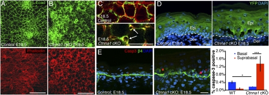
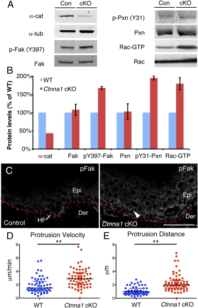
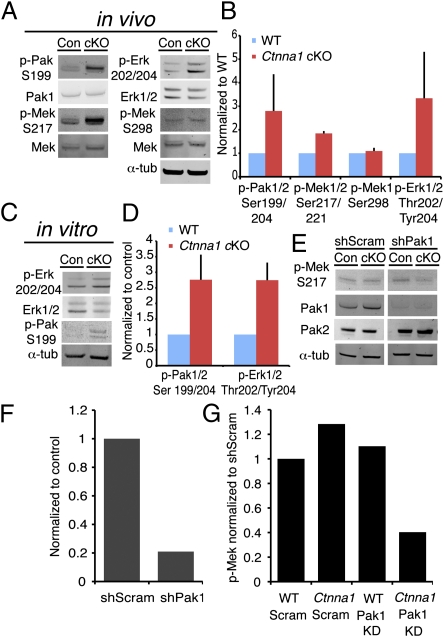
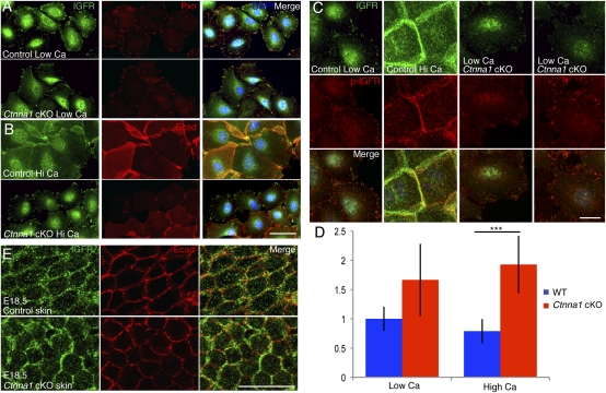
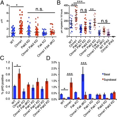
Cadherin/catenin-based adhesions coordinate cellular growth, survival, migration, and differentiation within a tissue by mechanically anchoring cells to their neighbors. They also intersect with diverse signaling pathways in development and cancer. Although the adhesive functions of adherens junction proteins are well characterized, their contribution to other signaling pathways is less well understood. Here, we show that ablation of α-catenin in the epidermis selectively induces apoptosis in suprabasal differentiating keratinocytes while sparing basal cell progenitors. This protection from death is coupled to elevated focal adhesion signaling, faster migration, and an altered distribution of growth factor receptors. We show that simultaneous depletion of α-catenin and focal adhesion kinase or p21-activated kinase eliminates basal cell protection as well as the elevated migration and proliferation of cells. The increased dependency of cells upon matrix interactions for their survival when cell-cell adhesions are destabilized has important implications for cancer progression and metastasis.


Conflict of interest statement

The authors declare no conflict of interest.

Figures

Fig. 1.
Fig. 1.
Morphological defects and suprabasal apoptosis in Ctnna1 cKO epidermis. (A and B) Projections of IFM confocal stacks of skin sections from E18.5 embryos, revealing disorganization within the 3D F-actin–AJ network of cKO epidermis. (C) Higher magnification of single E18.5 confocal sections revealing intercellular gaps within the cKO epidermal sheets (arrows). (D) Sagittal sections of E18.5 skins showing that cKO ruptures concentrate at the basal–suprabasal interface. YFP marks LV-Cre–infected cells. (E) Representative image and quantification of apoptotic cells in sagittal sections identified by active Caspase-3 immunostaining (red). Integrin β4 marks the base of cells within the basal layer. Error bars are ±SD. DAPI marks nuclei. [Scale bars (A and B): 50 μm; (D and E): 20 μm.]
Fig. 2.
Fig. 2.
Activation of focal adhesion signaling in the absence of α-catenin. (A) Immunoblots of in vivo skin lysates to assess the activation states of FAK, Paxillin, and Rac1. (B) Quantifications of data from A. (C) IFM reveals elevated activated (phosphorylated) FAK (arrowhead) at the leading front of the hair placode in control tissue and throughout the basal layer of Ctnna1 KO epidermis. (D and E) Quantifications of protrusion velocity (D) and protrusion distance (E) of keratinocytes migrating out of control and Ctnna1 cKO explants, as visualized by videomicroscopy over a 10-min interval. Black lines mark median ± interquartile range. Asterisks denote P < 0.01.
Fig. 3.
Fig. 3.
Increased Pak activation in Ctnna1 cKO epidermis promotes Erk/MAPK phosphorylation. (AE) Quantitative immunoblots of Pak1/2, Mek, and Erk1/2 phosphorylation levels from epidermal lysates from control and Ctnna1 cKO E18.5 embryos (A and B) and from their cultured keratinocytes (C and D). (E) Immunoblots of Mek phosphorylation upon in utero lentiviral transduction of Pak1 or scrambled shRNAs. (F) Quantifications of Pak1 knockdown efficiency. (G) Quantifications of relative Mek activity (phosphorylation) upon Pak1 knockdown.
Fig. 4.
Fig. 4.
Loss of junctional IGFR in Ctnna1-null keratinocytes. (A and B). Keratinocytes from WT and Ctnna1 cKO embryos were cultured in either low- or high-calcium medium and immunolabeled for IGFR, Paxillin, and/or E-cadherin as indicated. (C and D) Representative IFM images and quantifications to determine relative phospho-IGFR and IGFR levels in WT and Ctnna1-null keratinocytes in low and high calcium. (E) Planar views of WT and Ctnna1 cKO epidermis after immunolabeling E18.5 embryos for IGFR and E-cadherin. [Scale bars (A and B): 50 μm; (C and E): 20 μm.]
Fig. 5.
Fig. 5.
In vivo depletion of FAK and Pak proteins rescues migratory, proliferative, and apoptotic phenotypes of Ctnna1 cKO epidermis. (A) Quantifications of membrane protrusion distances in keratinocytes emerging from skin explants. Explants were imaged by videomicroscopy over 10 min. (B) Quantifications of keratinocyte outgrowth distances after 18 h of culturing explants from the embryos indicated and treated as indicated. (A and B) Black lines mark median ± interquartile range. ***P < 0.001; **P < 0.01; *P < 0.05; n.s., no significant difference. (C and D) Frequencies of phospho-histone H3-positive (mitotic) basal epidermal cells (C) and phospho-caspase-3–positive (apoptotic) basal versus suprabasal epidermal cells (D) from WT, cKO, and/or KD embryos as indicated. Quantification is based on tissues from more than two animals and >15 tissue sections/animal. Error bars represent ±SD.

References

    1. Halbleib JM, Nelson WJ. Cadherins in development: Cell adhesion, sorting, and tissue morphogenesis. Genes Dev. 2006;20:3199–3214. - PubMed
    1. Nelson WJ. Regulation of cell-cell adhesion by the cadherin-catenin complex. Biochem Soc Trans. 2008;36(2):149–155. - PMC - PubMed
    1. Tinkle CL, Pasolli HA, Stokes N, Fuchs E. New insights into cadherin function in epidermal sheet formation and maintenance of tissue integrity. Proc Natl Acad Sci USA. 2008;105:15405–15410. - PMC - PubMed
    1. Vasioukhin V, Bauer C, Degenstein L, Wise B, Fuchs E. Hyperproliferation and defects in epithelial polarity upon conditional ablation of alpha-catenin in skin. Cell. 2001;104:605–617. - PubMed
    1. Beronja S, Livshits G, Williams S, Fuchs E. Rapid functional dissection of genetic networks via tissue-specific transduction and RNAi in mouse embryos. Nat Med. 2010;16:821–827. - PMC - PubMed

Publication types

MeSH terms

Substances