

Twin-arginine-dependent translocation of folded proteins
- PMID: 22411976
- PMCID: PMC3297433
- DOI: 10.1098/rstb.2011.0202
Twin-arginine-dependent translocation of folded proteins
Erratum in
- Philos Trans R Soc Lond B Biol Sci. 2012 Aug 5;367(1599):2246
Abstract
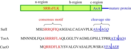
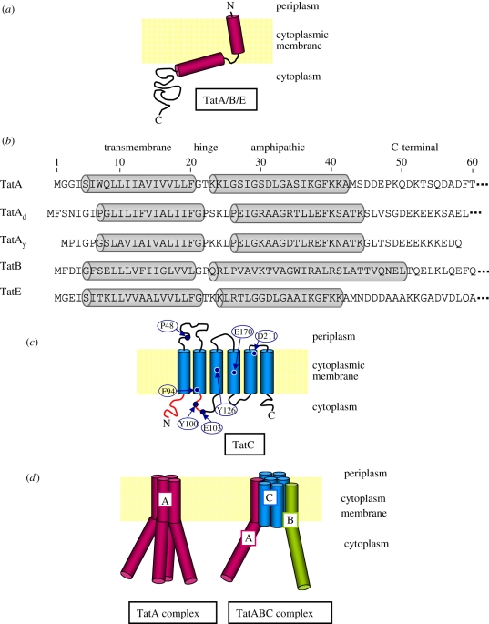
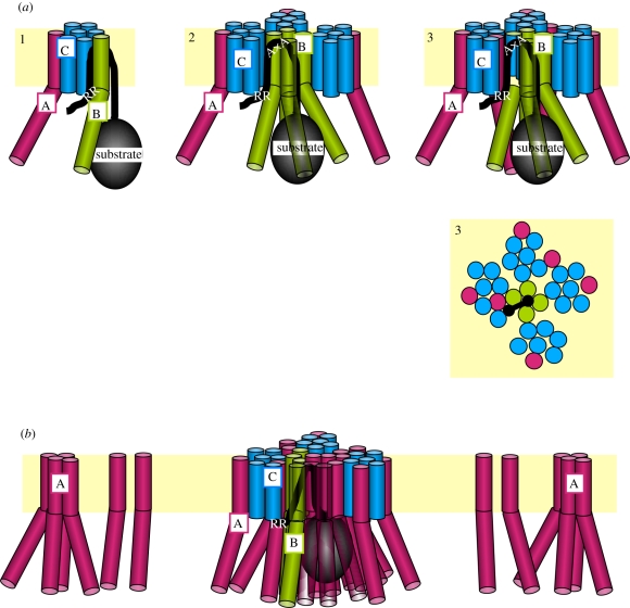
Twin-arginine translocation (Tat) denotes a protein transport pathway in bacteria, archaea and plant chloroplasts, which is specific for precursor proteins harbouring a characteristic twin-arginine pair in their signal sequences. Many Tat substrates receive cofactors and fold prior to translocation. For a subset of them, proofreading chaperones coordinate maturation and membrane-targeting. Tat translocases comprise two kinds of membrane proteins, a hexahelical TatC-type protein and one or two members of the single-spanning TatA protein family, called TatA and TatB. TatC- and TatA-type proteins form homo- and hetero-oligomeric complexes. The subunits of TatABC translocases are predominantly recovered from two separate complexes, a TatBC complex that might contain some TatA, and a homomeric TatA complex. TatB and TatC coordinately recognize twin-arginine signal peptides and accommodate them in membrane-embedded binding pockets. Advanced binding of the signal sequence to the Tat translocase requires the proton-motive force (PMF) across the membranes and might involve a first recruitment of TatA. When targeted in this manner, folded twin-arginine precursors induce homo-oligomerization of TatB and TatA. Ultimately, this leads to the formation of a transmembrane protein conduit that possibly consists of a pore-like TatA structure. The translocation step again is dependent on the PMF.
Figures
References
-
- Lee P. A., Tullman-Ercek D., Georgiou G. 2006. The bacterial twin-arginine translocation pathway. Annu. Rev. Microbiol. 60, 373–395 10.1146/annurev.micro.60.080805.142212 (doi:10.1146/annurev.micro.60.080805.142212) - DOI - PMC - PubMed
-
- Widdick D. A., Eijlander R. T., van Dijl J. M., Kuipers O. P., Palmer T. 2008. A facile reporter system for the experimental identification of twin-arginine translocation (Tat) signal peptides from all kingdoms of life. J. Mol. Biol. 375, 595–603 10.1016/j.jmb.2007.11.002 (doi:10.1016/j.jmb.2007.11.002) - DOI - PubMed
-
- Palmer T., Sargent F., Berks B. C. 2010. The Tat protein export pathway. In EcoSal—Escherichia coli and Salmonella: cellular and molecular biology (eds Böck A., Curtiss R., III, Kaper J. B., Karp P. D., Neidhardt F. C., Nyström T., Slauch J. M., Squires C. L., Ussery D.). Washington, DC: ASM Press
-
- Berks B. C. 1996. A common export pathway for proteins binding complex redox cofactors? Mol. Microbiol. 22, 393–404 10.1046/j.1365-2958.1996.00114.x (doi:10.1046/j.1365-2958.1996.00114.x) - DOI - PubMed
-
- Robinson C., Matos C. F. R. O., Beck D., Ren C., Lawrence J., Vasisht N., Mendel S. 2011. Transport and proofreading of proteins by the twin-arginine translocation (Tat) system in bacteria. Biochim. Biophys. Acta 1808, 876–884 10.1016/j.bbamem.2010.11.023 (doi:10.1016/j.bbamem.2010.11.023) - DOI - PubMed
Publication types
MeSH terms
Substances
LinkOut - more resources
Full Text Sources
Molecular Biology Databases