Review
. 2012 Apr 19;367(1592):1140-54.
doi: 10.1098/rstb.2011.0205.

The blueprint of the type-3 injectisome

Affiliations
Review

The blueprint of the type-3 injectisome

Agata Kosarewicz et al. Philos Trans R Soc Lond B Biol Sci. .

Abstract

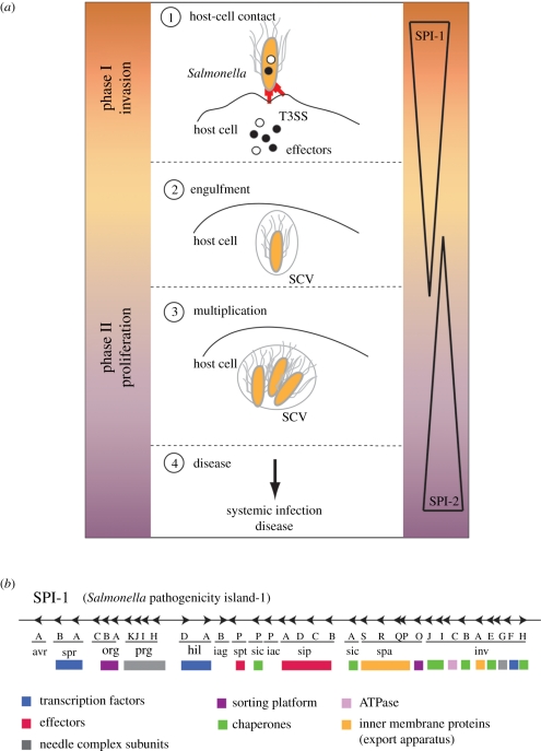
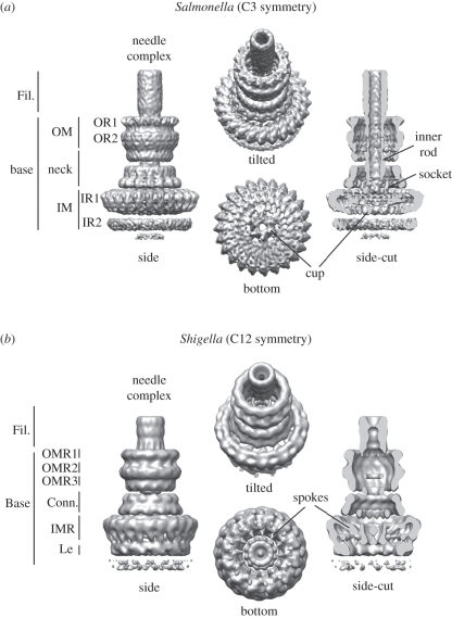
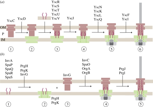
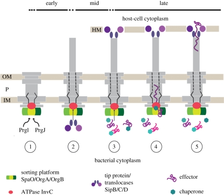
Type-3 secretion systems are sophisticated syringe-like nanomachines present in many animal and plant Gram-negative pathogens. They are capable of translocating an arsenal of specific bacterial toxins (effector proteins) from the prokaryotic cytoplasm across the three biological membranes directly into the eukaryotic cytosol, some of which modulate host cell mechanisms for the benefit of the pathogen. They populate a particular biological niche, which is maintained by specific, pathogen-dependent effectors. In contrast, the needle complex, which is the central component of this specialized protein delivery machine, is structurally well-conserved. It is a large supramolecular cylindrical structure composed of multiple copies of a relatively small subset of proteins, is embedded in the bacterial membranes and protrudes from the pathogen's surface with a needle filament. A central channel traverses the entire needle complex, and serves as a hollow conduit for proteins destined to travel this secretion pathway. In the past few years, there has been a tremendous increase in an understanding on both the structural and the mechanistic level. This review will thus focus on new insights of this remarkable molecular machine.


Figures

Figure 1.
Figure 1.
Infection pathway and organization of the T3SS. (a) Salmonella invasion scheme: (1) during the invasion phase (controlled by the Salmonella pathogenicity island 1, SPI-1), a contact between Salmonella and the host cell activates the SPI-1-encoded T3SS, which starts to secrete effector proteins that modify the cytoskeleton of the cell. This results in membrane ruffling that facilitates the subsequent uptake of the bacterium and formation of the Salmonella-containing vacuoles (SCVs) (2). (3) In the proliferation phase (maintained by SPI-2), the bacterium multiplies inside the SCVs before it is finally released to cause systemic infection (4). (b) Genomic organization of the Salmonella pathogenicity island-1 (SPI-1) and functional grouping of proteins: transcription factors, effectors, needle complex subunits, sorting platform, chaperones, inner membrane proteins (export apparatus), type-3 specific ATPase.
Figure 2.
Figure 2.
Needle complex structure from (a) Salmonella and (b) Shigella. Fil., needle filament; OM, outer membrane; OR, outer ring; OMR, outer membrane ring; IM, inner membrane; IR, inner ring; IMR, inner membrane ring; Conn., connector; Le, leg. Accession code: EMD-1875 (Salmonella), EMD-1617 (Shigella).
Figure 3.
Figure 3.
Domain structures of some needle complex proteins. (Summary of T3SS structures solved by X-ray, NMR, EM listed in table 2.) (a) Ribbon diagrams of domains of needle complex proteins building up the membrane-embedded base: InvG homologue EscC (X-ray, PDB: 3GR5), PrgK homologue EscJ (X-ray, PDB: 1YJ7), PrgH (C-terminal, periplasmic domain, X-ray, PDB: 3GR0), PrgH homologue MxiG (N-terminal, cytoplasmic domain, NMR, PDB: 2XXS). (OM (outer membrane), IM (inner membrane)). The periplasmic domains are organized into small modules. Domains D1 and D2 were speculated to be conserved ring-building motifs. (b) (Left panel) Crystal packing of PrgK homologue EscJ in a superhelix covering 24 subunits per turn (yellow). (Right panel) The modelled PrgK ring (yellow) based on the crystal contacts of EscJ but without helical rise. It displays the same diameters (approx. 180 Å) as the smaller ring of the inner ring obtained by selective disassembly of needle complexes. (c) Ribbon diagrams of domains of the needle filament forming proteins: PrgI from Salmonella (left) and MxiH in Shigella (right) (both X-ray, PDB 2X9C, 2CA5).
Figure 4.
Figure 4.
The structure of the needle complex at subnanometre resolution and docking of individual proteins. (a) Three-dimensional surface view of the Salmonella's needle complex. As different symmetries are present in the IR1 (24-fold) and OM/neck region (15-fold), symmetrization led to higher resolutions. (b) InvG (orange), PrgH (blue) and PrgK (yellow) were independently docked into the three-dimensional cryo-electron microscopy map of the needle complex as rigid bodies guided by their domain structure and secondary elements. (c) Fifteen monomers of InvG and 24 of PrgH/PrgK were docked into the lower neck ring and IR1, respectively. PrgH and PrgK are organized as concentric rings with different diameters. (d) A slice through the needle complex (side view) with docked atomic structures shows the organization of all three proteins—InvG, PrgH and PrgK—within the needle complex. Tube-like densities, which most probably correspond to the TM helices of PrgH and PrgK, connect IR1 with IR2 and define the localization of the inner membrane bilayer. Accession code: EMD-1874 and PDB 2Y9J (IR1), EMD-1871 and PDB 2Y9K (OM neck), EMD-1875 (needle complex)
Figure 5.
Figure 5.
Models of sequential needle complex assembly: (a) outside-in model (Yersinia) (b) inside-out model (Salmonella). OM, outer membrane; P, periplasm; IM, inner membrane.
Figure 6.
Figure 6.
Hierarchy and timing of substrate secretion. (1) In the early phase, PrgI and PrgJ are translocated to build up the inner rod and needle filament; (2) then the translocases are secreted to form the tip (SipD) at the distal end of the needle filament, which connects to the host cell membrane-embedded translocon (SipB, SipC) and establishes a physical connection between the bacterium and the host cell. (3) The effector proteins (late substrates) are translocated from the bacterial to the eukaryotic cytoplasm. Assisted by their cognate chaperones, they interact with the sorting platform (SpaO, OrgA and OrgB) and are then specifically recognized by the T3-associated ATPase (InvC). (4) ATP binding and hydrolysis are required to release the chaperone from its substrate and to unfold the substrate. The unfolded substrate is then believed to travel the secretion pathway through the inner rod and the needle filament (5) into the host cell cytoplasm. OM, outer membrane; P, periplasm; IM, inner membrane; HM, host cell membrane.

References

    1. Coburn B., Sekirov I., Finlay B. B. 2007. Type III secretion systems and disease. Clin. Microbiol. Rev. 20, 535–54910.1128/CMR.00013-07 (doi:10.1128/CMR.00013-07) - DOI - DOI - PMC - PubMed
    1. Galan J. E. 2001. Salmonella interactions with host cells: type III secretion at work. Annu. Rev. Cell Dev. Biol. 17, 53–8610.1146/annurev.cellbio.17.1.53 (doi:10.1146/annurev.cellbio.17.1.53) - DOI - DOI - PubMed
    1. Akopyan K., Edgren T., Wang-Edgren H., Rosqvist R., Fahlgren A., Wolf-Watz H., Fallman M. 2011. Translocation of surface-localized effectors in type III secretion. Proc. Natl Acad. Sci. USA 108, 1639–164410.1073/pnas.1013888108 (doi:10.1073/pnas.1013888108) - DOI - DOI - PMC - PubMed
    1. Galan J. E., Wolf-Watz H. 2006. Protein delivery into eukaryotic cells by type III secretion machines. Nature 444, 567–57310.1038/nature05272 (doi:10.1038/nature05272) - DOI - DOI - PubMed
    1. Hueck C. J. 1998. Type III protein secretion systems in bacterial pathogens of animals and plants. Microbiol. Mol. Biol. Rev. 62, 379–433 - PMC - PubMed

Publication types

MeSH terms

LinkOut - more resources